 ムービー
ムービー コントローラー


+ データを開く

- 基本情報
基本情報
| 登録情報 | データベース: EMDB / ID: EMD-23968 | |||||||||
|---|---|---|---|---|---|---|---|---|---|---|
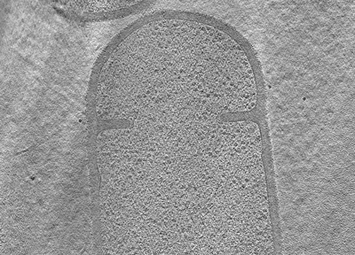
| タイトル | Tomogram of a dividing sporulating cell of Bacillus subtilis SpoIIE null mutant (Figure 8A of the manuscript Khanna et al., 2001) | |||||||||
 マップデータ マップデータ | Tomogram of a dividing sporulating cell of Bacillus subtilis SpoIIE null mutant | |||||||||
 試料 試料 |
| |||||||||
| 生物種 |  | |||||||||
| 手法 | 電子線トモグラフィー法 / クライオ電子顕微鏡法 | |||||||||
 データ登録者 データ登録者 | Khanna K / Villa E | |||||||||
| 資金援助 |  米国, 2件 米国, 2件
| |||||||||
 引用 引用 | ジャーナル: Elife / 年: 2021 タイトル: Asymmetric localization of the cell division machinery during sporulation. 著者: Kanika Khanna / Javier Lopez-Garrido / Joseph Sugie / Kit Pogliano / Elizabeth Villa / 要旨: The Gram-positive bacterium can divide via two modes. During vegetative growth, the division septum is formed at the midcell to produce two equal daughter cells. However, during sporulation, the ...The Gram-positive bacterium can divide via two modes. During vegetative growth, the division septum is formed at the midcell to produce two equal daughter cells. However, during sporulation, the division septum is formed closer to one pole to yield a smaller forespore and a larger mother cell. Using cryo-electron tomography, genetics and fluorescence microscopy, we found that the organization of the division machinery is different in the two septa. While FtsAZ filaments, the major orchestrators of bacterial cell division, are present uniformly around the leading edge of the invaginating vegetative septa, they are only present on the mother cell side of the invaginating sporulation septa. We provide evidence suggesting that the different distribution and number of FtsAZ filaments impact septal thickness, causing vegetative septa to be thicker than sporulation septa already during constriction. Finally, we show that a sporulation-specific protein, SpoIIE, regulates asymmetric divisome localization and septal thickness during sporulation. | |||||||||
| 履歴 |
|
- 構造の表示
構造の表示
| ムービー |
 ムービービューア ムービービューア |
|---|---|
| 添付画像 |
- ダウンロードとリンク
-EMDBアーカイブ
| マップデータ | emd_23968.map.gz | 302.2 MB | EMDBマップデータ形式 | |
|---|---|---|---|---|
| ヘッダ (付随情報) | emd-23968-v30.xmlemd-23968.xml | 9.2 KB 9.2 KB | 表示 表示 | EMDBヘッダ |
| 画像 |  emd_23968.png emd_23968.png | 183 KB | ||
| アーカイブディレクトリ |  http://ftp.pdbj.org/pub/emdb/structures/EMD-23968ftp://ftp.pdbj.org/pub/emdb/structures/EMD-23968 http://ftp.pdbj.org/pub/emdb/structures/EMD-23968ftp://ftp.pdbj.org/pub/emdb/structures/EMD-23968 | HTTPS FTP |
-検証レポート
| 文書・要旨 | emd_23968_validation.pdf.gz | 281.1 KB | 表示 | EMDB検証レポート |
|---|---|---|---|---|
| 文書・詳細版 | emd_23968_full_validation.pdf.gz | 280.6 KB | 表示 | |
| XML形式データ | emd_23968_validation.xml.gz | 4.8 KB | 表示 | |
| アーカイブディレクトリ | https://ftp.pdbj.org/pub/emdb/validation_reports/EMD-23968ftp://ftp.pdbj.org/pub/emdb/validation_reports/EMD-23968 | HTTPS FTP |
-関連構造データ
| 関連構造データ | C: 同じ文献を引用 ( |
|---|---|
| 電子顕微鏡画像生データ | EMPIAR-10710 (タイトル: Tilt series of dividing vegetative and sporulating cells of Bacillus subtilis from the manuscript - Khanna et al., 2021 Data size: 8.7 Data #1: Tilt series of a dividing vegetative cell of Bacillus subtilis (Figure 3A of the manuscript Khanna et al., 2021) [tilt series] Data #2: Tomogram of a dividing vegetative cell of Bacillus subtilis (Figure 4A of the manuscript Khanna et al., 2021) [tilt series] Data #3: Tomogram of a dividing vegetative cell of Bacillus subtilis FtsZ-linker(Q-rich) strain (Figure 5A and 6A of the manuscript Khanna et al., 2021) [tilt series] Data #4: Tomogram of a dividing sporulating cell of Bacillus subtilis (Figure 7A of the manuscript Khanna et al., 2021) [tilt series] Data #5: Tomogram of a dividing sporulating cell of Bacillus subtilis (Figure 7 - figure supplement 4 of the manuscript Khanna et al., 2021) [tilt series] Data #6: Tomogram of a dividing sporulating cell of Bacillus subtilis SpoIIE null mutant (Figure 8A of the manuscript Khanna et al., 2001) [tilt series]) |
-リンク
| EMDBのページ | EMDB (EBI/PDBe) / EMDataResource |
|---|
-マップ
| ファイル | ダウンロード / ファイル: emd_23968.map.gz / 形式: CCP4 / 大きさ: 424.8 MB / タイプ: IMAGE STORED AS SIGNED BYTE | ||||||||||||||||||||||||||||||||||||||||||||||||||||||||||||||||||||
|---|---|---|---|---|---|---|---|---|---|---|---|---|---|---|---|---|---|---|---|---|---|---|---|---|---|---|---|---|---|---|---|---|---|---|---|---|---|---|---|---|---|---|---|---|---|---|---|---|---|---|---|---|---|---|---|---|---|---|---|---|---|---|---|---|---|---|---|---|---|
| 注釈 | Tomogram of a dividing sporulating cell of Bacillus subtilis SpoIIE null mutant | ||||||||||||||||||||||||||||||||||||||||||||||||||||||||||||||||||||
| ボクセルのサイズ | X=Y=Z: 21.356 Å | ||||||||||||||||||||||||||||||||||||||||||||||||||||||||||||||||||||
| 密度 |
| ||||||||||||||||||||||||||||||||||||||||||||||||||||||||||||||||||||
| 対称性 | 空間群: 1 | ||||||||||||||||||||||||||||||||||||||||||||||||||||||||||||||||||||
| 詳細 | EMDB XML:
CCP4マップ ヘッダ情報:
| ||||||||||||||||||||||||||||||||||||||||||||||||||||||||||||||||||||
-添付データ
- 試料の構成要素
-全体 : Dividing sporulating cell of Bacillus subtilis SpoIIE null mutant
| 全体 | 名称: Dividing sporulating cell of Bacillus subtilis SpoIIE null mutant |
|---|---|
| 要素 |
|
-超分子 #1: Dividing sporulating cell of Bacillus subtilis SpoIIE null mutant
| 超分子 | 名称: Dividing sporulating cell of Bacillus subtilis SpoIIE null mutant タイプ: cell / ID: 1 / 親要素: 0 / 詳細: Figure 8A of the manuscript Khanna et al., 2021 |
|---|---|
| 由来(天然) | 生物種: |
-実験情報
-構造解析
| 手法 | クライオ電子顕微鏡法 |
|---|---|
 解析 解析 | 電子線トモグラフィー法 |
| 試料の集合状態 | cell |
-試料調製
| 緩衝液 | pH: 7 |
|---|---|
| 凍結 | 凍結剤: ETHANE-PROPANE |
| 切片作成 | 集束イオンビーム - 装置: OTHER / 集束イオンビーム - イオン: OTHER / 集束イオンビーム - 電圧: 5 kV / 集束イオンビーム - 電流: 0.05 nA / 集束イオンビーム - 時間: 1800 sec. / 集束イオンビーム - 温度: 93 K / 集束イオンビーム - Initial thickness: 1200 nm / 集束イオンビーム - 最終 厚さ: 200 nm 集束イオンビーム - 詳細: The value given for _emd_sectioning_focused_ion_beam.instrument is Thermo Scientific Aquilos. This is not in a list of allowed values {'DB235', 'OTHER'} so OTHER is ...集束イオンビーム - 詳細: The value given for _emd_sectioning_focused_ion_beam.instrument is Thermo Scientific Aquilos. This is not in a list of allowed values {'DB235', 'OTHER'} so OTHER is written into the XML file. |
- 電子顕微鏡法
| 顕微鏡 | FEI TITAN KRIOS |
|---|---|
| 撮影 | フィルム・検出器のモデル: GATAN K2 SUMMIT (4k x 4k) 平均電子線量: 1.0 e/Å2 |
| 電子線 | 加速電圧: 300 kV / 電子線源:  FIELD EMISSION GUN FIELD EMISSION GUN |
| 電子光学系 | 照射モード: FLOOD BEAM / 撮影モード: BRIGHT FIELD |
| 実験機器 |  モデル: Titan Krios / 画像提供: FEI Company |
-画像解析
| 最終 再構成 | ソフトウェア - 名称: IMOD / 使用した粒子像数: 59 |
|---|
