+ Open data
- Basic information
| Entry | Database: EMDB / ID: EMD-2391 | |||||||||
|---|---|---|---|---|---|---|---|---|---|---|
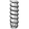
| Title | Human Respiratory Syncytial Virus ribonucleocapsid | |||||||||
 Map data Map data | Subtomogram average of hRSV nucleocapsid | |||||||||
 Sample Sample |
| |||||||||
 Keywords Keywords | RSV / RNP / nucleocapsid | |||||||||
| Biological species |  Human respiratory syncytial virus Human respiratory syncytial virus | |||||||||
| Method | subtomogram averaging / cryo EM / Resolution: 40.0 Å | |||||||||
 Authors Authors | Liljeroos L / Krzyzaniak MA / Helenius A / Butcher SJ | |||||||||
 Citation Citation |  Journal: Proc Natl Acad Sci U S A / Year: 2013 Journal: Proc Natl Acad Sci U S A / Year: 2013Title: Architecture of respiratory syncytial virus revealed by electron cryotomography. Authors: Lassi Liljeroos / Magdalena Anna Krzyzaniak / Ari Helenius / Sarah Jane Butcher /  Abstract: Human respiratory syncytial virus is a human pathogen that causes severe infection of the respiratory tract. Current information about the structure of the virus and its interaction with host cells ...Human respiratory syncytial virus is a human pathogen that causes severe infection of the respiratory tract. Current information about the structure of the virus and its interaction with host cells is limited. We carried out an electron cryotomographic characterization of cell culture-grown human respiratory syncytial virus to determine the architecture of the virion. The particles ranged from 100 nm to 1,000 nm in diameter and were spherical, filamentous, or a combination of the two. The filamentous morphology correlated with the presence of a cylindrical matrix protein layer linked to the inner leaflet of the viral envelope and with local ordering of the glycoprotein spikes. Recombinant viruses with only the fusion protein in their envelope showed that these glycoproteins were predominantly in the postfusion conformation, but some were also in the prefusion form. The ribonucleocapsids were left-handed, randomly oriented, and curved inside the virions. In filamentous particles, they were often adjacent to an intermediate layer of protein assigned to M2-1 (an envelope-associated protein known to mediate association of ribonucleocapsids with the matrix protein). Our results indicate important differences in structure between the Paramyxovirinae and Pneumovirinae subfamilies within the Paramyxoviridae, and provide fresh insights into host cell exit of a serious pathogen. | |||||||||
| History |
|
- Structure visualization
| Movie |
 Movie viewer Movie viewer |
|---|---|
| Structure viewer | EM map:  SurfView SurfView Molmil Molmil Jmol/JSmol Jmol/JSmol |
| Supplemental images |
- Downloads & links
-EMDB archive
| Map data |  emd_2391.map.gz emd_2391.map.gz | 953.3 KB |  EMDB map data format EMDB map data format | |
|---|---|---|---|---|
| Header (meta data) |  emd-2391-v30.xml emd-2391-v30.xml emd-2391.xml emd-2391.xml | 8.6 KB 8.6 KB | Display Display |  EMDB header EMDB header |
| Images |  emd_2391.tif emd_2391.tif | 55.5 KB | ||
| Archive directory |  http://ftp.pdbj.org/pub/emdb/structures/EMD-2391 http://ftp.pdbj.org/pub/emdb/structures/EMD-2391 ftp://ftp.pdbj.org/pub/emdb/structures/EMD-2391 ftp://ftp.pdbj.org/pub/emdb/structures/EMD-2391 | HTTPS FTP |
-Validation report
| Summary document |  emd_2391_validation.pdf.gz emd_2391_validation.pdf.gz | 203.7 KB | Display |  EMDB validaton report EMDB validaton report |
|---|---|---|---|---|
| Full document |  emd_2391_full_validation.pdf.gz emd_2391_full_validation.pdf.gz | 202.7 KB | Display | |
| Data in XML |  emd_2391_validation.xml.gz emd_2391_validation.xml.gz | 5.2 KB | Display | |
| Arichive directory |  https://ftp.pdbj.org/pub/emdb/validation_reports/EMD-2391 https://ftp.pdbj.org/pub/emdb/validation_reports/EMD-2391 ftp://ftp.pdbj.org/pub/emdb/validation_reports/EMD-2391 ftp://ftp.pdbj.org/pub/emdb/validation_reports/EMD-2391 | HTTPS FTP |
-Related structure data
- Links
Links
| EMDB pages |  EMDB (EBI/PDBe) / EMDB (EBI/PDBe) /  EMDataResource EMDataResource |
|---|
- Map
Map
| File |  Download / File: emd_2391.map.gz / Format: CCP4 / Size: 1001 KB / Type: IMAGE STORED AS FLOATING POINT NUMBER (4 BYTES) Download / File: emd_2391.map.gz / Format: CCP4 / Size: 1001 KB / Type: IMAGE STORED AS FLOATING POINT NUMBER (4 BYTES) | ||||||||||||||||||||||||||||||||||||||||||||||||||||||||||||||||||||
|---|---|---|---|---|---|---|---|---|---|---|---|---|---|---|---|---|---|---|---|---|---|---|---|---|---|---|---|---|---|---|---|---|---|---|---|---|---|---|---|---|---|---|---|---|---|---|---|---|---|---|---|---|---|---|---|---|---|---|---|---|---|---|---|---|---|---|---|---|---|
| Annotation | Subtomogram average of hRSV nucleocapsid | ||||||||||||||||||||||||||||||||||||||||||||||||||||||||||||||||||||
| Projections & slices | Image control
Images are generated by Spider. | ||||||||||||||||||||||||||||||||||||||||||||||||||||||||||||||||||||
| Voxel size | X=Y=Z: 7.68 Å | ||||||||||||||||||||||||||||||||||||||||||||||||||||||||||||||||||||
| Density |
| ||||||||||||||||||||||||||||||||||||||||||||||||||||||||||||||||||||
| Symmetry | Space group: 1 | ||||||||||||||||||||||||||||||||||||||||||||||||||||||||||||||||||||
| Details | EMDB XML:
CCP4 map header:
| ||||||||||||||||||||||||||||||||||||||||||||||||||||||||||||||||||||
-Supplemental data
- Sample components
-Entire : Human Respiratory Syncytial Virus ribonucleocapsid
| Entire | Name: Human Respiratory Syncytial Virus ribonucleocapsid |
|---|---|
| Components |
|
-Supramolecule #1000: Human Respiratory Syncytial Virus ribonucleocapsid
| Supramolecule | Name: Human Respiratory Syncytial Virus ribonucleocapsid / type: sample / ID: 1000 / Number unique components: 1 |
|---|
-Supramolecule #1: Human respiratory syncytial virus
| Supramolecule | Name: Human respiratory syncytial virus / type: virus / ID: 1 / Name.synonym: ribonucleocapsid / NCBI-ID: 11250 / Sci species name: Human respiratory syncytial virus / Database: NCBI / Virus type: VIRION / Virus isolate: SPECIES / Virus enveloped: No / Virus empty: Yes / Syn species name: ribonucleocapsid |
|---|---|
| Host (natural) | Organism:  Homo sapiens (human) / synonym: VERTEBRATES Homo sapiens (human) / synonym: VERTEBRATES |
-Experimental details
-Structure determination
| Method | cryo EM |
|---|---|
 Processing Processing | subtomogram averaging |
| Aggregation state | particle |
- Sample preparation
| Buffer | pH: 7.4 / Details: HBSS, 25 mM Hepes |
|---|---|
| Grid | Details: C-flat 2/2-2C and C-flat 2/2-4C, holey carbon copper grid |
| Vitrification | Cryogen name: ETHANE / Instrument: HOMEMADE PLUNGER / Details: Vitrification instrument: Homemade plunger / Method: Blot for 4 seconds before plunging |
- Electron microscopy
| Microscope | FEI TECNAI F20 |
|---|---|
| Date | Jul 11, 2012 |
| Image recording | Category: CCD / Film or detector model: GATAN ULTRASCAN 4000 (4k x 4k) |
| Electron beam | Acceleration voltage: 200 kV / Electron source:  FIELD EMISSION GUN FIELD EMISSION GUN |
| Electron optics | Calibrated magnification: 39400 / Illumination mode: FLOOD BEAM / Imaging mode: BRIGHT FIELD / Cs: 2.0 mm / Nominal defocus max: 6.0 µm / Nominal defocus min: 6.0 µm / Nominal magnification: 39400 |
| Sample stage | Specimen holder: Eucentric / Specimen holder model: GATAN LIQUID NITROGEN / Tilt series - Axis1 - Min angle: -60 ° / Tilt series - Axis1 - Max angle: 60 ° |
| Experimental equipment |  Model: Tecnai F20 / Image courtesy: FEI Company |
- Image processing
| Details | Subtomograms were manually selected from the tomograms by visual inspection |
|---|---|
| Final reconstruction | Resolution.type: BY AUTHOR / Resolution: 40.0 Å / Resolution method: FSC 0.5 CUT-OFF / Software - Name: IMOD, PEET, Bsoft / Number subtomograms used: 1402 |
 Movie
Movie Controller


 Z (Sec.)
 Y (Row.)
 X (Col.)


