 Movie
Movie Controller

[English] 日本語
 Yorodumi
- EMDB-23863: Cryo-EM structure of SidJ-SdeC-CaM reaction intermediate complex -
+ Open data

- Basic information
| Entry | Database: EMDB / ID: EMD-23863 | ||||||||||||
|---|---|---|---|---|---|---|---|---|---|---|---|---|---|
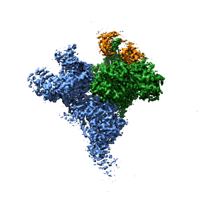
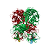
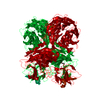
| Title | Cryo-EM structure of SidJ-SdeC-CaM reaction intermediate complex | ||||||||||||
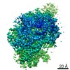
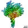
 Map data Map data | |||||||||||||
 Sample Sample |
| ||||||||||||
 Keywords Keywords | SidJ / SdeC / CaM / complex / Intermediate / Acyl / Adenylate / Legionella / Ubiquitination / TRANSFERASE / TRANSFERASE-LIGASE complex | ||||||||||||
| Function / homology |  Function and homology information Function and homology informationprotein-glutamic acid ligase activity, initiating / protein-glutamic acid ligase activity, elongating / Ligases / NAD+-protein-arginine ADP-ribosyltransferase activity / transporter inhibitor activity / negative regulation of ryanodine-sensitive calcium-release channel activity / negative regulation of calcium ion export across plasma membrane / presynaptic endocytosis / calcineurin-mediated signaling / regulation of cell communication by electrical coupling involved in cardiac conduction ...protein-glutamic acid ligase activity, initiating / protein-glutamic acid ligase activity, elongating / Ligases / NAD+-protein-arginine ADP-ribosyltransferase activity / transporter inhibitor activity / negative regulation of ryanodine-sensitive calcium-release channel activity / negative regulation of calcium ion export across plasma membrane / presynaptic endocytosis / calcineurin-mediated signaling / regulation of cell communication by electrical coupling involved in cardiac conduction / adenylate cyclase binding / protein phosphatase activator activity / protein deubiquitination / detection of calcium ion / regulation of cardiac muscle contraction / catalytic complex / calcium channel inhibitor activity / presynaptic cytosol / regulation of release of sequestered calcium ion into cytosol by sarcoplasmic reticulum / titin binding / regulation of cardiac muscle contraction by regulation of the release of sequestered calcium ion / regulation of calcium-mediated signaling / cysteine-type peptidase activity / voltage-gated potassium channel complex / calcium channel complex / substantia nigra development / regulation of heart rate / calyx of Held / adenylate cyclase activator activity / regulation of cytokinesis / protein serine/threonine kinase activator activity / spindle microtubule / sarcomere / calcium channel regulator activity / response to calcium ion / G2/M transition of mitotic cell cycle / spindle pole / calcium-dependent protein binding / long-term synaptic potentiation / myelin sheath / synaptic vesicle membrane / transferase activity / sperm midpiece / vesicle / transmembrane transporter binding / G protein-coupled receptor signaling pathway / nucleotide binding / calcium ion binding / centrosome / protein kinase binding / protein-containing complex / proteolysis / membrane / metal ion binding / nucleus / plasma membrane / cytoplasm Similarity search - Function | ||||||||||||
| Biological species |   Legionella pneumophila (bacteria) / Legionella pneumophila (bacteria) /  Homo sapiens (human) Homo sapiens (human) | ||||||||||||
| Method | single particle reconstruction / cryo EM / Resolution: 2.8 Å | ||||||||||||
 Authors Authors | Osinski A / Black MH | ||||||||||||
| Funding support |  United States, 3 items United States, 3 items
| ||||||||||||
 Citation Citation | Journal: Mol Cell / Year: 2021 Title: Structural and mechanistic basis for protein glutamylation by the kinase fold. Authors: Adam Osinski / Miles H Black / Krzysztof Pawłowski / Zhe Chen / Yang Li / Vincent S Tagliabracci /  Abstract: The kinase domain transfers phosphate from ATP to substrates. However, the Legionella effector SidJ adopts a kinase fold, yet catalyzes calmodulin (CaM)-dependent glutamylation to inactivate the SidE ...The kinase domain transfers phosphate from ATP to substrates. However, the Legionella effector SidJ adopts a kinase fold, yet catalyzes calmodulin (CaM)-dependent glutamylation to inactivate the SidE ubiquitin ligases. The structural and mechanistic basis in which the kinase domain catalyzes protein glutamylation is unknown. Here we present cryo-EM reconstructions of SidJ:CaM:SidE reaction intermediate complexes. We show that the kinase-like active site of SidJ adenylates an active-site Glu in SidE, resulting in the formation of a stable reaction intermediate complex. An insertion in the catalytic loop of the kinase domain positions the donor Glu near the acyl-adenylate for peptide bond formation. Our structural analysis led us to discover that the SidJ paralog SdjA is a glutamylase that differentially regulates the SidE ligases during Legionella infection. Our results uncover the structural and mechanistic basis in which the kinase fold catalyzes non-ribosomal amino acid ligations and reveal an unappreciated level of SidE-family regulation. | ||||||||||||
| History |
|
- Structure visualization
| Movie |
Movie viewer |
|---|---|
| Structure viewer | EM map: SurfViewMolmilJmol/JSmol |
| Supplemental images |
- Downloads & links
-EMDB archive
| Map data | emd_23863.map.gz | 228.9 MB | EMDB map data format | |
|---|---|---|---|---|
| Header (meta data) | emd-23863-v30.xmlemd-23863.xml | 19.9 KB 19.9 KB | Display Display | EMDB header |
| Images |  emd_23863.png emd_23863.png | 95.2 KB | ||
| Filedesc metadata | emd-23863.cif.gz | 7.5 KB | ||
| Archive directory |  http://ftp.pdbj.org/pub/emdb/structures/EMD-23863ftp://ftp.pdbj.org/pub/emdb/structures/EMD-23863 http://ftp.pdbj.org/pub/emdb/structures/EMD-23863ftp://ftp.pdbj.org/pub/emdb/structures/EMD-23863 | HTTPS FTP |
-Related structure data
| Related structure data |  7misMC  7mirC M: atomic model generated by this map C: citing same article ( |
|---|---|
| Similar structure data |


-Links
| EMDB pages | EMDB (EBI/PDBe) / EMDataResource |
|---|---|
| Related items in Molecule of the Month |

-Map
| File | Download / File: emd_23863.map.gz / Format: CCP4 / Size: 244.1 MB / Type: IMAGE STORED AS FLOATING POINT NUMBER (4 BYTES) | ||||||||||||||||||||||||||||||||||||||||||||||||||||||||||||||||||||
|---|---|---|---|---|---|---|---|---|---|---|---|---|---|---|---|---|---|---|---|---|---|---|---|---|---|---|---|---|---|---|---|---|---|---|---|---|---|---|---|---|---|---|---|---|---|---|---|---|---|---|---|---|---|---|---|---|---|---|---|---|---|---|---|---|---|---|---|---|---|
| Projections & slices | Image control
Images are generated by Spider. | ||||||||||||||||||||||||||||||||||||||||||||||||||||||||||||||||||||
| Voxel size | X=Y=Z: 1.08 Å | ||||||||||||||||||||||||||||||||||||||||||||||||||||||||||||||||||||
| Density |
| ||||||||||||||||||||||||||||||||||||||||||||||||||||||||||||||||||||
| Symmetry | Space group: 1 | ||||||||||||||||||||||||||||||||||||||||||||||||||||||||||||||||||||
| Details | EMDB XML:
CCP4 map header:
| ||||||||||||||||||||||||||||||||||||||||||||||||||||||||||||||||||||
 Z (Sec.)
 Y (Row.)
 X (Col.)


-Supplemental data
- Sample components
+Entire : Cryo-EM structure of SidJ-SdeC-CaM reaction intermediate complex
+Supramolecule #1: Cryo-EM structure of SidJ-SdeC-CaM reaction intermediate complex
+Supramolecule #2: SidJ-SdeC
+Supramolecule #3: Calmodulin
+Macromolecule #1: Calmodulin-dependent glutamylase SidJ
+Macromolecule #2: Calmodulin
+Macromolecule #3: SdeC
+Macromolecule #4: ADENOSINE-5'-TRIPHOSPHATE

+Macromolecule #5: MAGNESIUM ION
+Macromolecule #6: SODIUM ION
+Macromolecule #7: CALCIUM ION
+Macromolecule #8: ADENOSINE MONOPHOSPHATE

-Experimental details
-Structure determination
| Method | cryo EM |
|---|---|
 Processing Processing | single particle reconstruction |
| Aggregation state | particle |
-Sample preparation
| Concentration | 0.35 mg/mL |
|---|---|
| Buffer | pH: 6.5 Details: 25 mM Bis-tris pH 6.5, 100 mM NaCl, 1 mM TCEP, 2 mM MgCl2, 1 mM ATP |
| Vitrification | Cryogen name: ETHANE / Instrument: FEI VITROBOT MARK IV |
- Electron microscopy
| Microscope | FEI TITAN KRIOS |
|---|---|
| Image recording | Film or detector model: GATAN K3 BIOQUANTUM (6k x 4k) / Average electron dose: 50.0 e/Å2 |
| Electron beam | Acceleration voltage: 300 kV / Electron source:  FIELD EMISSION GUN FIELD EMISSION GUN |
| Electron optics | Illumination mode: FLOOD BEAM / Imaging mode: BRIGHT FIELD |
| Experimental equipment |  Model: Titan Krios / Image courtesy: FEI Company |
