 Movie
Movie Controller


+ Open data

- Basic information
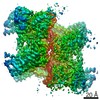
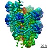
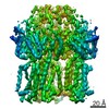
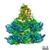
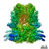
| Entry | Database: EMDB / ID: EMD-23720 | |||||||||
|---|---|---|---|---|---|---|---|---|---|---|
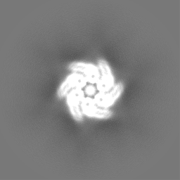
| Title | ATPgS bound TnsC filament from shCAST system | |||||||||
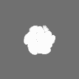
 Map data Map data | Postprocessed map | |||||||||
 Sample Sample |
| |||||||||
 Keywords Keywords | CRISPR / Transposition / AAA+ ATPase / Tn7 / DNA BINDING PROTEIN-DNA complex | |||||||||
| Biological species |  Scytonema hofmannii (bacteria) / synthetic construct (others) Scytonema hofmannii (bacteria) / synthetic construct (others) | |||||||||
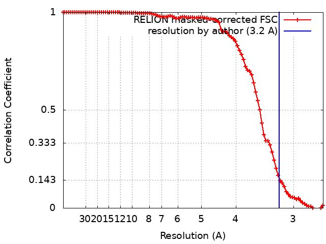
| Method | helical reconstruction / cryo EM / Resolution: 3.2 Å | |||||||||
 Authors Authors | Park J / Tsai AWL | |||||||||
| Funding support |  United States, 1 items United States, 1 items
| |||||||||
 Citation Citation | Journal: Science / Year: 2021 Title: Structural basis for target site selection in RNA-guided DNA transposition systems. Authors: Jung-Un Park / Amy Wei-Lun Tsai / Eshan Mehrotra / Michael T Petassi / Shan-Chi Hsieh / Ailong Ke / Joseph E Peters / Elizabeth H Kellogg / Abstract: CRISPR-associated transposition systems allow guide RNA-directed integration of a single DNA cargo in one orientation at a fixed distance from a programmable target sequence. We used cryo-electron ...CRISPR-associated transposition systems allow guide RNA-directed integration of a single DNA cargo in one orientation at a fixed distance from a programmable target sequence. We used cryo-electron microscopy (cryo-EM) to define the mechanism that underlies this process by characterizing the transposition regulator, TnsC, from a type V-K CRISPR-transposase system. In this scenario, polymerization of adenosine triphosphate-bound TnsC helical filaments could explain how polarity information is passed to the transposase. TniQ caps the TnsC filament, representing a universal mechanism for target information transfer in Tn7/Tn7-like elements. Transposase-driven disassembly establishes delivery of the element only to unused protospacers. Finally, TnsC transitions to define the fixed point of insertion, as revealed by structures with the transition state mimic ADP•AlF These mechanistic findings provide the underpinnings for engineering CRISPR-associated transposition systems for research and therapeutic applications. | |||||||||
| History |
|
- Structure visualization
| Movie |
 Movie viewer Movie viewer |
|---|---|
| Structure viewer | EM map: SurfViewMolmilJmol/JSmol |
| Supplemental images |

- Downloads & links
-EMDB archive
| Map data | emd_23720.map.gz | 5.7 MB | EMDB map data format | |
|---|---|---|---|---|
| Header (meta data) | emd-23720-v30.xmlemd-23720.xml | 25.6 KB 25.6 KB | Display Display | EMDB header |
| FSC (resolution estimation) | emd_23720_fsc.xml | 9.1 KB | Display | FSC data file |
| Images | 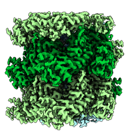 emd_23720.png emd_23720.png | 113.7 KB | ||
| Masks | emd_23720_msk_1.map | 64 MB | Mask map | |
| Filedesc metadata | emd-23720.cif.gz | 6.9 KB | ||
| Others | emd_23720_additional_1.map.gzemd_23720_half_map_1.map.gzemd_23720_half_map_2.map.gz | 49.2 MB 49.5 MB 49.5 MB | ||
| Archive directory |  http://ftp.pdbj.org/pub/emdb/structures/EMD-23720ftp://ftp.pdbj.org/pub/emdb/structures/EMD-23720 http://ftp.pdbj.org/pub/emdb/structures/EMD-23720ftp://ftp.pdbj.org/pub/emdb/structures/EMD-23720 | HTTPS FTP |
-Related structure data
| Related structure data | 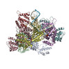 7m99MC  7m9aC  7m9bC  7m9cC  7n6iC C: citing same article ( M: atomic model generated by this map |
|---|---|
| Similar structure data |


-Links
| EMDB pages | EMDB (EBI/PDBe) / EMDataResource |
|---|
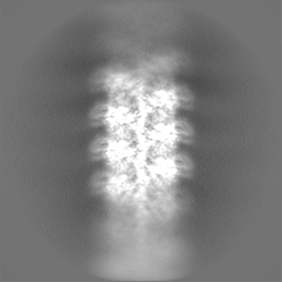
-Map
| File | Download / File: emd_23720.map.gz / Format: CCP4 / Size: 64 MB / Type: IMAGE STORED AS FLOATING POINT NUMBER (4 BYTES) | ||||||||||||||||||||||||||||||||||||||||||||||||||||||||||||||||||||
|---|---|---|---|---|---|---|---|---|---|---|---|---|---|---|---|---|---|---|---|---|---|---|---|---|---|---|---|---|---|---|---|---|---|---|---|---|---|---|---|---|---|---|---|---|---|---|---|---|---|---|---|---|---|---|---|---|---|---|---|---|---|---|---|---|---|---|---|---|---|
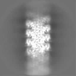
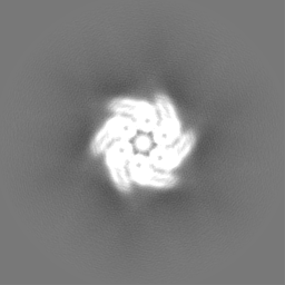
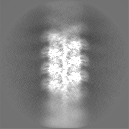
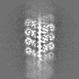
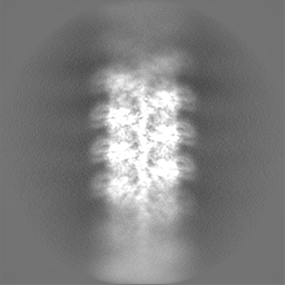
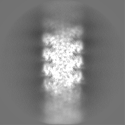
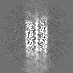
| Annotation | Postprocessed map | ||||||||||||||||||||||||||||||||||||||||||||||||||||||||||||||||||||
| Projections & slices | Image control
Images are generated by Spider. | ||||||||||||||||||||||||||||||||||||||||||||||||||||||||||||||||||||
| Voxel size | X=Y=Z: 1.33 Å | ||||||||||||||||||||||||||||||||||||||||||||||||||||||||||||||||||||
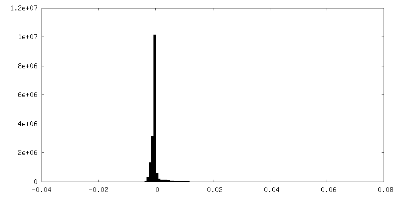
| Density |
| ||||||||||||||||||||||||||||||||||||||||||||||||||||||||||||||||||||
| Symmetry | Space group: 1 | ||||||||||||||||||||||||||||||||||||||||||||||||||||||||||||||||||||
| Details | EMDB XML:
CCP4 map header:
| ||||||||||||||||||||||||||||||||||||||||||||||||||||||||||||||||||||
 Z (Sec.)
 Y (Row.)
 X (Col.)


-Supplemental data
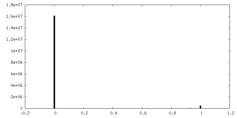
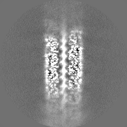
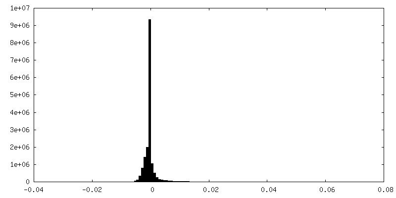
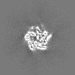
-Mask #1
| File | emd_23720_msk_1.map | ||||||||||||
|---|---|---|---|---|---|---|---|---|---|---|---|---|---|
| Projections & Slices |
| ||||||||||||
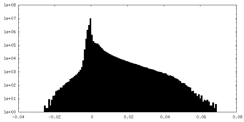
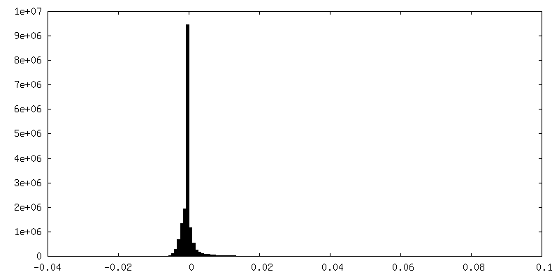
| Density Histograms |


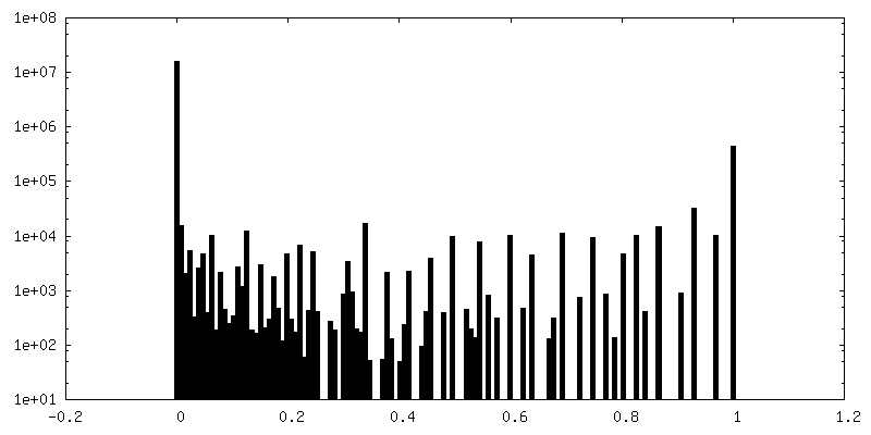
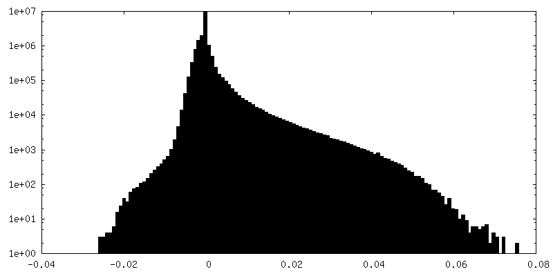
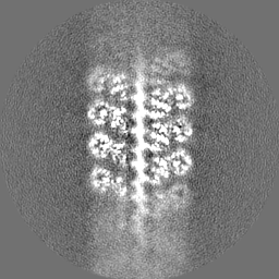
-Additional map: Full map
| File | emd_23720_additional_1.map | ||||||||||||
|---|---|---|---|---|---|---|---|---|---|---|---|---|---|
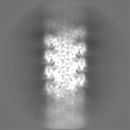
| Annotation | Full map | ||||||||||||
| Projections & Slices |
| ||||||||||||
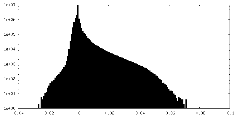
| Density Histograms |


-Half map: Even half-map
| File | emd_23720_half_map_1.map | ||||||||||||
|---|---|---|---|---|---|---|---|---|---|---|---|---|---|
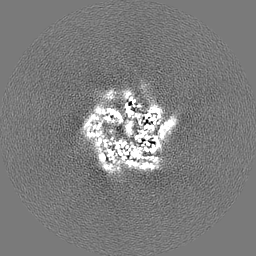
| Annotation | Even half-map | ||||||||||||
| Projections & Slices |
| ||||||||||||
| Density Histograms |

-Half map: Odd half-map
| File | emd_23720_half_map_2.map | ||||||||||||
|---|---|---|---|---|---|---|---|---|---|---|---|---|---|
| Annotation | Odd half-map | ||||||||||||
| Projections & Slices |
| ||||||||||||
| Density Histograms |
- Sample components
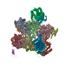
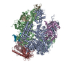
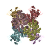
-Entire : Helical filament of TnsC monomers bound to DNA
| Entire | Name: Helical filament of TnsC monomers bound to DNA |
|---|---|
| Components |
|
-Supramolecule #1: Helical filament of TnsC monomers bound to DNA
| Supramolecule | Name: Helical filament of TnsC monomers bound to DNA / type: complex / ID: 1 / Parent: 0 / Macromolecule list: #1-#3 / Details: Reconstituted with ATPgS |
|---|---|
| Source (natural) | Organism: Scytonema hofmannii (bacteria) |
| Molecular weight | Theoretical: 45.4 kDa/nm |
-Macromolecule #1: TnsC
| Macromolecule | Name: TnsC / type: protein_or_peptide / ID: 1 / Number of copies: 8 / Enantiomer: LEVO |
|---|---|
| Source (natural) | Organism: Scytonema hofmannii (bacteria) |
| Molecular weight | Theoretical: 31.444617 KDa |
| Recombinant expression | Organism: |
| Sequence | String: MTEAQAIAKQ LGGVKPDDEW LQAEIARLKG KSIVPLQQVK TLHDWLDGKR KARKSCRVVG ESRTGKTVAC DAYRYRHKPQ QEAGRPPTV PVVYIRPHQK CGPKDLFKKI TEYLKYRVTK GTVSDFRDRT IEVLKGCGVE MLIIDEADRL KPETFADVRD I AEDLGIAV ...String: MTEAQAIAKQ LGGVKPDDEW LQAEIARLKG KSIVPLQQVK TLHDWLDGKR KARKSCRVVG ESRTGKTVAC DAYRYRHKPQ QEAGRPPTV PVVYIRPHQK CGPKDLFKKI TEYLKYRVTK GTVSDFRDRT IEVLKGCGVE MLIIDEADRL KPETFADVRD I AEDLGIAV VLVGTDRLDA VIKRDEQVLE RFRAHLRFGK LSGEDFKNTV EMWEQMVLKL PVSSNLKSKE MLRILTSATE GY IGRLDEI LREAAIRSLS RGLKKIDKAV LQEVAKEYK |
-Macromolecule #2: DNA (5'-D(P*TP*TP*TP*TP*TP*TP*TP*TP*TP*TP*TP*TP*TP*TP*TP*TP*TP*TP...
| Macromolecule | Name: DNA (5'-D(P*TP*TP*TP*TP*TP*TP*TP*TP*TP*TP*TP*TP*TP*TP*TP*TP*TP*TP*T)-3') type: dna / ID: 2 / Number of copies: 1 / Classification: DNA |
|---|---|
| Source (natural) | Organism: synthetic construct (others) |
| Molecular weight | Theoretical: 6.343091 KDa |
| Sequence | String: (DT)(DT)(DT)(DT)(DT)(DT)(DT)(DT)(DT)(DT) (DT)(DT)(DT)(DT)(DT)(DT)(DT)(DT)(DT)(DT) (DT) |
-Macromolecule #3: DNA (5'-D(P*AP*AP*AP*AP*AP*AP*AP*AP*AP*AP*AP*AP*AP*AP*AP*AP*AP*AP...
| Macromolecule | Name: DNA (5'-D(P*AP*AP*AP*AP*AP*AP*AP*AP*AP*AP*AP*AP*AP*AP*AP*AP*AP*AP*A)-3') type: dna / ID: 3 / Number of copies: 1 / Classification: DNA |
|---|---|
| Source (natural) | Organism: synthetic construct (others) |
| Molecular weight | Theoretical: 6.532386 KDa |
| Sequence | String: (DA)(DA)(DA)(DA)(DA)(DA)(DA)(DA)(DA)(DA) (DA)(DA)(DA)(DA)(DA)(DA)(DA)(DA)(DA)(DA) (DA) |
-Macromolecule #4: PHOSPHOTHIOPHOSPHORIC ACID-ADENYLATE ESTER
| Macromolecule | Name: PHOSPHOTHIOPHOSPHORIC ACID-ADENYLATE ESTER / type: ligand / ID: 4 / Number of copies: 7 / Formula: AGS |
|---|---|
| Molecular weight | Theoretical: 523.247 Da |
| Chemical component information |  ChemComp-AGS: |
-Macromolecule #5: MAGNESIUM ION
| Macromolecule | Name: MAGNESIUM ION / type: ligand / ID: 5 / Number of copies: 7 / Formula: MG |
|---|---|
| Molecular weight | Theoretical: 24.305 Da |
-Experimental details
-Structure determination
| Method | cryo EM |
|---|---|
 Processing Processing | helical reconstruction |
| Aggregation state | filament |
-Sample preparation
| Concentration | 1 mg/mL | |||||||||||||||||||||
|---|---|---|---|---|---|---|---|---|---|---|---|---|---|---|---|---|---|---|---|---|---|---|
| Buffer | pH: 7.4 Component:
Details: Sample was diluted into low salt using dialysis. | |||||||||||||||||||||
| Grid | Model: Quantifoil R1.2/1.3 / Material: GOLD / Mesh: 300 / Support film - Material: GOLD / Support film - topology: HOLEY / Support film - Film thickness: 50 / Pretreatment - Type: GLOW DISCHARGE / Pretreatment - Time: 30 sec. / Pretreatment - Atmosphere: AIR / Pretreatment - Pressure: 0.038 kPa Details: Grid cleaned with solvent and examined for defects under light microscope. | |||||||||||||||||||||
| Vitrification | Cryogen name: ETHANE / Chamber humidity: 100 % / Chamber temperature: 277.15 K / Instrument: FEI VITROBOT MARK IV / Details: Blot for 6 seconds before plunging. | |||||||||||||||||||||
| Details | This sample comprised monodisperse filaments reconstituted using dialysis. |
- Electron microscopy
| Microscope | FEI TALOS ARCTICA |
|---|---|
| Temperature | Min: 93.15 K / Max: 103.2 K |
| Image recording | Film or detector model: GATAN K3 BIOQUANTUM (6k x 4k) / Digitization - Dimensions - Width: 5760 pixel / Digitization - Dimensions - Height: 4092 pixel / Number grids imaged: 1 / Number real images: 3474 / Average exposure time: 3.0 sec. / Average electron dose: 40.0 e/Å2 |
| Electron beam | Acceleration voltage: 200 kV / Electron source:  FIELD EMISSION GUN FIELD EMISSION GUN |
| Electron optics | C2 aperture diameter: 50.0 µm / Illumination mode: FLOOD BEAM / Imaging mode: BRIGHT FIELD / Cs: 2.7 mm / Nominal defocus max: 2.5 µm / Nominal defocus min: 1.0 µm / Nominal magnification: 63000 |
| Sample stage | Specimen holder model: FEI TITAN KRIOS AUTOGRID HOLDER / Cooling holder cryogen: NITROGEN |
| Experimental equipment |  Model: Talos Arctica / Image courtesy: FEI Company |
+Image processing
-Atomic model buiding 1
| Refinement | Space: REAL / Protocol: AB INITIO MODEL / Overall B value: 50 / Target criteria: Correlation coefficient |
|---|---|
| Output model | PDB-7m99: |
