ムービー
ムービー コントローラー


+ データを開く

- 基本情報
基本情報
| 登録情報 | データベース: EMDB / ID: EMD-2365 | |||||||||
|---|---|---|---|---|---|---|---|---|---|---|
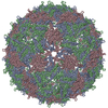
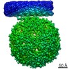
| タイトル | Asymmetric structure of a virus-receptor complex | |||||||||
 マップデータ マップデータ | Sub-tomogram average of bacteriophage MS2 bound to its receptor | |||||||||
 試料 試料 |
| |||||||||
 キーワード キーワード | virus / receptor / complex / asymmetric / bacteriophage | |||||||||
| 機能・相同性 |  機能・相同性情報 機能・相同性情報negative regulation of viral translation / T=3 icosahedral viral capsid / regulation of translation / structural molecule activity / RNA binding / identical protein binding 類似検索 - 分子機能 | |||||||||
| 生物種 |   Enterobacterio phage MS2 (ファージ) Enterobacterio phage MS2 (ファージ) | |||||||||
| 手法 | サブトモグラム平均法 / クライオ電子顕微鏡法 / 解像度: 39.0 Å | |||||||||
 データ登録者 データ登録者 | Dent KC / Thompson R / Barker AM / Barr JN / Hiscox JA / Stockley PG / Ranson NA | |||||||||
 引用 引用 | ジャーナル: Structure / 年: 2013 タイトル: The asymmetric structure of an icosahedral virus bound to its receptor suggests a mechanism for genome release. 著者: Kyle C Dent / Rebecca Thompson / Amy M Barker / Julian A Hiscox / John N Barr / Peter G Stockley / Neil A Ranson /  要旨: Simple, spherical RNA viruses have well-understood, symmetric protein capsids, but little structural information is available for their asymmetric components, such as minor proteins and their ...Simple, spherical RNA viruses have well-understood, symmetric protein capsids, but little structural information is available for their asymmetric components, such as minor proteins and their genomes, which are vital for infection. Here, we report an asymmetric structure of bacteriophage MS2, attached to its receptor, the F-pilus. Cryo-electron tomography and subtomographic averaging of such complexes result in a structure containing clear density for the packaged genome, implying that the conformation of the genome is the same in each virus particle. The data also suggest that the single-copy viral maturation protein breaks the symmetry of the capsid, occupying a position that would be filled by a coat protein dimer in an icosahedral shell. This capsomere can thus fulfill its known biological roles in receptor and genome binding and suggests an exit route for the genome during infection. | |||||||||
| 履歴 |
|
- 構造の表示
構造の表示
| ムービー |
ムービービューア |
|---|---|
| 構造ビューア | EMマップ: SurfViewMolmilJmol/JSmol |
| 添付画像 |
 UCSF Chimera
- ダウンロードとリンク
-EMDBアーカイブ
| マップデータ | emd_2365.map.gz | 895.9 KB | EMDBマップデータ形式 | |
|---|---|---|---|---|
| ヘッダ (付随情報) | emd-2365-v30.xmlemd-2365.xml | 9.5 KB 9.5 KB | 表示 表示 | EMDBヘッダ |
| 画像 | 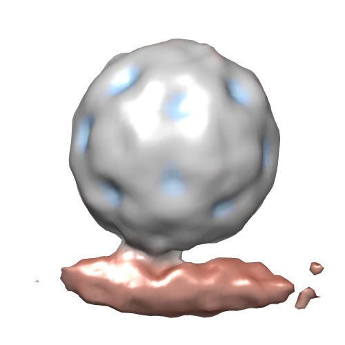 emd_2365.png emd_2365.png | 78.6 KB | ||
| アーカイブディレクトリ |  http://ftp.pdbj.org/pub/emdb/structures/EMD-2365ftp://ftp.pdbj.org/pub/emdb/structures/EMD-2365 http://ftp.pdbj.org/pub/emdb/structures/EMD-2365ftp://ftp.pdbj.org/pub/emdb/structures/EMD-2365 | HTTPS FTP |
-検証レポート
| 文書・要旨 | emd_2365_validation.pdf.gz | 219.2 KB | 表示 | EMDB検証レポート |
|---|---|---|---|---|
| 文書・詳細版 | emd_2365_full_validation.pdf.gz | 218.3 KB | 表示 | |
| XML形式データ | emd_2365_validation.xml.gz | 5.1 KB | 表示 | |
| アーカイブディレクトリ | https://ftp.pdbj.org/pub/emdb/validation_reports/EMD-2365ftp://ftp.pdbj.org/pub/emdb/validation_reports/EMD-2365 | HTTPS FTP |
-関連構造データ


-リンク
| EMDBのページ | EMDB (EBI/PDBe) / EMDataResource |
|---|---|
| 「今月の分子」の関連する項目 |

-マップ
| ファイル | ダウンロード / ファイル: emd_2365.map.gz / 形式: CCP4 / 大きさ: 1001 KB / タイプ: IMAGE STORED AS FLOATING POINT NUMBER (4 BYTES) | ||||||||||||||||||||||||||||||||||||||||||||||||||||||||||||||||||||
|---|---|---|---|---|---|---|---|---|---|---|---|---|---|---|---|---|---|---|---|---|---|---|---|---|---|---|---|---|---|---|---|---|---|---|---|---|---|---|---|---|---|---|---|---|---|---|---|---|---|---|---|---|---|---|---|---|---|---|---|---|---|---|---|---|---|---|---|---|---|
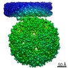
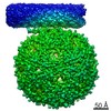
| 注釈 | Sub-tomogram average of bacteriophage MS2 bound to its receptor | ||||||||||||||||||||||||||||||||||||||||||||||||||||||||||||||||||||
| 投影像・断面図 | 画像のコントロール
画像は Spider により作成 | ||||||||||||||||||||||||||||||||||||||||||||||||||||||||||||||||||||
| ボクセルのサイズ | X=Y=Z: 9.12 Å | ||||||||||||||||||||||||||||||||||||||||||||||||||||||||||||||||||||
| 密度 |
| ||||||||||||||||||||||||||||||||||||||||||||||||||||||||||||||||||||
| 対称性 | 空間群: 1 | ||||||||||||||||||||||||||||||||||||||||||||||||||||||||||||||||||||
| 詳細 | EMDB XML:
CCP4マップ ヘッダ情報:
| ||||||||||||||||||||||||||||||||||||||||||||||||||||||||||||||||||||
 Z (Sec.)
 Y (Row.)
 X (Col.)


-添付データ
- 試料の構成要素
-全体 : Bacteriophage MS2 bound to its receptor, the E. coli F-pilus
| 全体 | 名称: Bacteriophage MS2 bound to its receptor, the E. coli F-pilus |
|---|---|
| 要素 |
|
-超分子 #1000: Bacteriophage MS2 bound to its receptor, the E. coli F-pilus
| 超分子 | 名称: Bacteriophage MS2 bound to its receptor, the E. coli F-pilus タイプ: sample / ID: 1000 / 詳細: Sub-tomographic averaging of virus-decorated pili 集合状態: One T=3 icosahedral virus bound to a pilus fibre Number unique components: 2 |
|---|
-超分子 #1: Enterobacterio phage MS2
| 超分子 | 名称: Enterobacterio phage MS2 / タイプ: virus / ID: 1 / NCBI-ID: 12022 / 生物種: Enterobacterio phage MS2 / ウイルスタイプ: VIRION / ウイルス・単離状態: SPECIES / ウイルス・エンベロープ: No / ウイルス・中空状態: No |
|---|---|
| 宿主 | 生物種: |
| ウイルス殻 | Shell ID: 1 / 直径: 290 Å / T番号(三角分割数): 3 |
-超分子 #2: F-Pilus
| 超分子 | 名称: F-Pilus / タイプ: organelle_or_cellular_component / ID: 2 / 組換発現: No |
|---|---|
| 由来(天然) | 生物種: |
-実験情報
-構造解析
| 手法 | クライオ電子顕微鏡法 |
|---|---|
 解析 解析 | サブトモグラム平均法 |
| 試料の集合状態 | particle |
-試料調製
| グリッド | 詳細: 200 mesh lacey carbon grids, glow discharged in air. |
|---|---|
| 凍結 | 凍結剤: ETHANE / 装置: HOMEMADE PLUNGER |
- 電子顕微鏡法
| 顕微鏡 | FEI TECNAI 12 |
|---|---|
| 日付 | 2012年1月1日 |
| 撮影 | カテゴリ: CCD フィルム・検出器のモデル: GATAN ULTRASCAN 1000 (2k x 2k) |
| 電子線 | 加速電圧: 120 kV / 電子線源: LAB6 |
| 電子光学系 | 倍率(補正後): 23000 / 照射モード: FLOOD BEAM / 撮影モード: BRIGHT FIELD / 最大 デフォーカス(公称値): 3.0 µm / 最小 デフォーカス(公称値): 3.0 µm / 倍率(公称値): 23000 |
| 試料ステージ | 試料ホルダーモデル: GATAN LIQUID NITROGEN / Tilt series - Axis1 - Min angle: -60 ° / Tilt series - Axis1 - Max angle: 60 ° |
-画像解析
| 最終 再構成 | 想定した対称性 - 点群: C1 (非対称) / 解像度のタイプ: BY AUTHOR / 解像度: 39.0 Å / 解像度の算出法: FSC 0.5 CUT-OFF / ソフトウェア - 名称: IMOD, PEET / 使用したサブトモグラム数: 1000 |
|---|