 Movie
Movie Controller

[English] 日本語
 Yorodumi
- EMDB-23610: Cryo-EM asymmetric structure of Grafix-stabilized PaFS octameric ... -
+ Open data

- Basic information
| Entry | Database: EMDB / ID: EMD-23610 | |||||||||
|---|---|---|---|---|---|---|---|---|---|---|
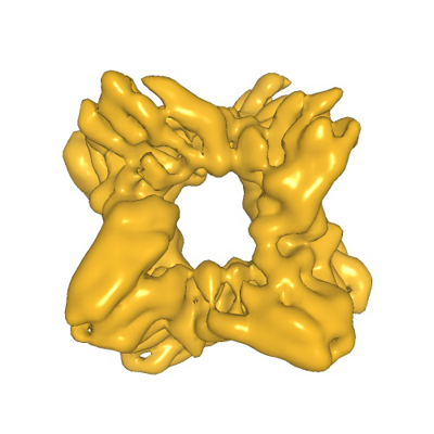
| Title | Cryo-EM asymmetric structure of Grafix-stabilized PaFS octameric prenyltransferase domain | |||||||||
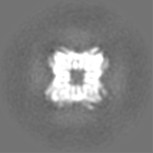
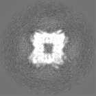
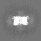
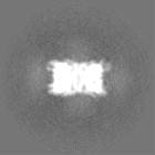
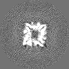
 Map data Map data | Octameric core prenyltransferase domain of PaFS, subjected to crosslinking, with imposed C2 symmetry | |||||||||
 Sample Sample |
| |||||||||
| Function / homology |  Function and homology information Function and homology informationfusicocca-2,10(14)-diene synthase / alcohol biosynthetic process / mycotoxin biosynthetic process / geranylgeranyl diphosphate synthase / geranylgeranyl diphosphate synthase activity / isoprenoid biosynthetic process / lyase activity / metal ion binding Similarity search - Function | |||||||||
| Biological species |  Diaporthe amygdali (fungus) Diaporthe amygdali (fungus) | |||||||||
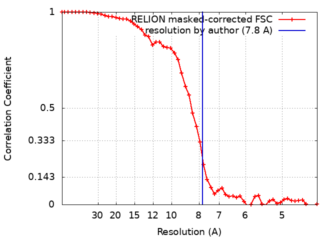
| Method | single particle reconstruction / cryo EM / Resolution: 7.8 Å | |||||||||
 Authors Authors | Faylo JL / van Eeuwen T / Murakami K / Christianson DW | |||||||||
| Funding support |  United States, 1 items United States, 1 items
| |||||||||
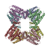
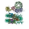
 Citation Citation | Journal: Nat Commun / Year: 2021 Title: Structural insight on assembly-line catalysis in terpene biosynthesis. Authors: Jacque L Faylo / Trevor van Eeuwen / Hee Jong Kim / Jose J Gorbea Colón / Benjamin A Garcia / Kenji Murakami / David W Christianson / Abstract: Fusicoccadiene synthase from Phomopsis amygdali (PaFS) is a unique bifunctional terpenoid synthase that catalyzes the first two steps in the biosynthesis of the diterpene glycoside Fusicoccin A, a ...Fusicoccadiene synthase from Phomopsis amygdali (PaFS) is a unique bifunctional terpenoid synthase that catalyzes the first two steps in the biosynthesis of the diterpene glycoside Fusicoccin A, a mediator of 14-3-3 protein interactions. The prenyltransferase domain of PaFS generates geranylgeranyl diphosphate, which the cyclase domain then utilizes to generate fusicoccadiene, the tricyclic hydrocarbon skeleton of Fusicoccin A. Here, we use cryo-electron microscopy to show that the structure of full-length PaFS consists of a central octameric core of prenyltransferase domains, with the eight cyclase domains radiating outward via flexible linker segments in variable splayed-out positions. Cryo-electron microscopy and chemical crosslinking experiments additionally show that compact conformations can be achieved in which cyclase domains are more closely associated with the prenyltransferase core. This structural analysis provides a framework for understanding substrate channeling, since most of the geranylgeranyl diphosphate generated by the prenyltransferase domains remains on the enzyme for cyclization to form fusicoccadiene. | |||||||||
| History |
|
- Structure visualization
| Movie |
Movie viewer |
|---|---|
| Structure viewer | EM map: SurfViewMolmilJmol/JSmol |
| Supplemental images |
- Downloads & links
-EMDB archive
| Map data | emd_23610.map.gz | 7.9 MB | EMDB map data format | |
|---|---|---|---|---|
| Header (meta data) | emd-23610-v30.xmlemd-23610.xml | 17.6 KB 17.6 KB | Display Display | EMDB header |
| FSC (resolution estimation) | emd_23610_fsc.xml | 5.1 KB | Display | FSC data file |
| Images |  emd_23610.png emd_23610.png | 118.6 KB | ||
| Masks | emd_23610_msk_1.map | 10.5 MB | Mask map | |
| Others | emd_23610_half_map_1.map.gzemd_23610_half_map_2.map.gz | 7.9 MB 7.9 MB | ||
| Archive directory |  http://ftp.pdbj.org/pub/emdb/structures/EMD-23610ftp://ftp.pdbj.org/pub/emdb/structures/EMD-23610 http://ftp.pdbj.org/pub/emdb/structures/EMD-23610ftp://ftp.pdbj.org/pub/emdb/structures/EMD-23610 | HTTPS FTP |
-Related structure data


-Links
| EMDB pages | EMDB (EBI/PDBe) / EMDataResource |
|---|---|
| Related items in Molecule of the Month |

-Map
| File | Download / File: emd_23610.map.gz / Format: CCP4 / Size: 10.5 MB / Type: IMAGE STORED AS FLOATING POINT NUMBER (4 BYTES) | ||||||||||||||||||||||||||||||||||||||||||||||||||||||||||||||||||||
|---|---|---|---|---|---|---|---|---|---|---|---|---|---|---|---|---|---|---|---|---|---|---|---|---|---|---|---|---|---|---|---|---|---|---|---|---|---|---|---|---|---|---|---|---|---|---|---|---|---|---|---|---|---|---|---|---|---|---|---|---|---|---|---|---|---|---|---|---|---|
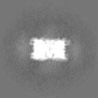
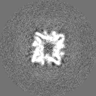
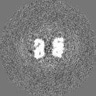
| Annotation | Octameric core prenyltransferase domain of PaFS, subjected to crosslinking, with imposed C2 symmetry | ||||||||||||||||||||||||||||||||||||||||||||||||||||||||||||||||||||
| Projections & slices | Image control
Images are generated by Spider. | ||||||||||||||||||||||||||||||||||||||||||||||||||||||||||||||||||||
| Voxel size | X=Y=Z: 2.16 Å | ||||||||||||||||||||||||||||||||||||||||||||||||||||||||||||||||||||
| Density |
| ||||||||||||||||||||||||||||||||||||||||||||||||||||||||||||||||||||
| Symmetry | Space group: 1 | ||||||||||||||||||||||||||||||||||||||||||||||||||||||||||||||||||||
| Details | EMDB XML:
CCP4 map header:
| ||||||||||||||||||||||||||||||||||||||||||||||||||||||||||||||||||||
 Z (Sec.)
 Y (Row.)
 X (Col.)


-Supplemental data
-Mask #1
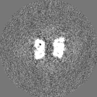
| File | emd_23610_msk_1.map | ||||||||||||
|---|---|---|---|---|---|---|---|---|---|---|---|---|---|
| Projections & Slices |
| ||||||||||||
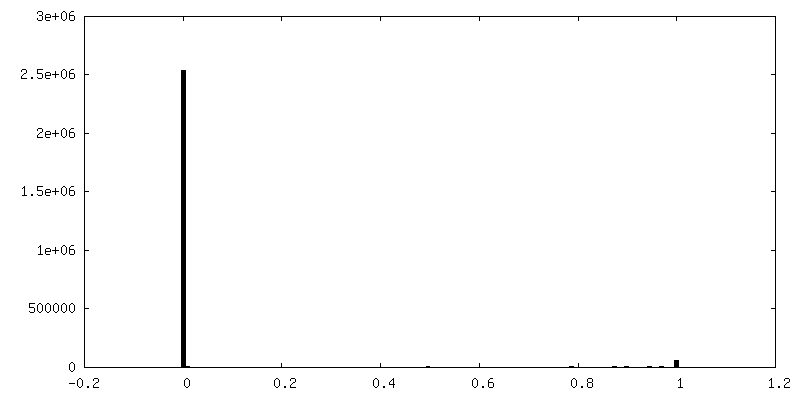
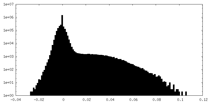
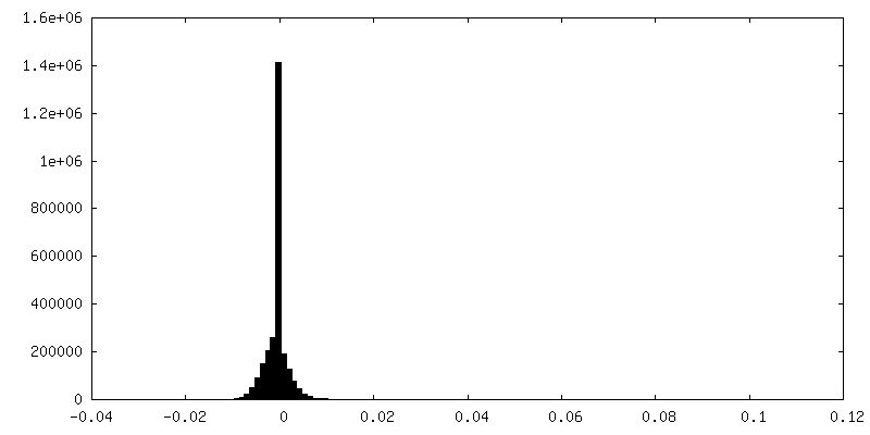
| Density Histograms |


-Half map: unsharpened, unfiltered half-map, particles subjected to partial signal...
| File | emd_23610_half_map_1.map | ||||||||||||
|---|---|---|---|---|---|---|---|---|---|---|---|---|---|
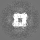
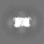
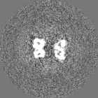
| Annotation | unsharpened, unfiltered half-map, particles subjected to partial signal subtraction | ||||||||||||
| Projections & Slices |
| ||||||||||||
| Density Histograms |


-Half map: unsharpened, unfiltered half-map, particles subjected to partial signal...
| File | emd_23610_half_map_2.map | ||||||||||||
|---|---|---|---|---|---|---|---|---|---|---|---|---|---|
| Annotation | unsharpened, unfiltered half-map, particles subjected to partial signal subtraction | ||||||||||||
| Projections & Slices |
| ||||||||||||
| Density Histograms |

- Sample components
-Entire : Apo octameric core prenyltransferase domain of PaFS
| Entire | Name: Apo octameric core prenyltransferase domain of PaFS |
|---|---|
| Components |
|
-Supramolecule #1: Apo octameric core prenyltransferase domain of PaFS
| Supramolecule | Name: Apo octameric core prenyltransferase domain of PaFS / type: complex / ID: 1 / Parent: 0 / Macromolecule list: all Details: Reconstruction of single domain of full-length complex was calculated by partial signal subtraction. Refinement imposed C2 symmetry. |
|---|---|
| Source (natural) | Organism: Diaporthe amygdali (fungus) |
| Recombinant expression | Organism:  |
| Molecular weight | Experimental: 392 KDa |
-Macromolecule #1: Fusicoccadiene synthase from Phomopsis amygdali
| Macromolecule | Name: Fusicoccadiene synthase from Phomopsis amygdali / type: protein_or_peptide / ID: 1 / Enantiomer: LEVO / EC number: fusicocca-2,10(14)-diene synthase |
|---|---|
| Source (natural) | Organism: Diaporthe amygdali (fungus) |
| Recombinant expression | Organism: |
| Sequence | String: MGSSHHHHHH SSGLVPRGSH MEFKYSEVVE PSTYYTEGLC EGIDVRKSKF TTLEDRGAIR AHEDWNKHIG PCGEYRGTLG PRFSFISVA VPECIPERLE VISYANEFAF LHDDVTDHVG HDTGEVENDE MMTVFLEAAH T GAIDTSNK VDIRRAGKKR IQSQLFLEML ...String: MGSSHHHHHH SSGLVPRGSH MEFKYSEVVE PSTYYTEGLC EGIDVRKSKF TTLEDRGAIR AHEDWNKHIG PCGEYRGTLG PRFSFISVA VPECIPERLE VISYANEFAF LHDDVTDHVG HDTGEVENDE MMTVFLEAAH T GAIDTSNK VDIRRAGKKR IQSQLFLEML AIDPECAKTT MKSWARFVEV GSSRQHETRF VE LAKYIPY RIMDVGEMFW FGLVTFGLGL HIPDHELELC RELMANAWIA VGLQNDIWSW PKE RDAATL HGKDHVVNAI WVLMQEHQTD VDGAMQICRK LIVEYVAKYL EVIEATKNDE SISL DLRKY LDAMLYSISG NVVWSLECPR YNPDVSFNKT QLEWMRQGLP SLESCPVLAR SPEID SDES AVSPTADESD STEDSLGSGS RQDSSLSTGL SLSPVHSNEG KDLQRVDTDH IFFEKA VLE APYDYIASMP SKGVRDQFID ALNDWLRVPD VKVGKIKDAV RVLHNSSLLL DDFQDNS PL RRGKPSTHNI FGSAQTVNTA TYSIIKAIGQ IMEFSAGESV QEVMNSIMIL FQGQAMDL F WTYNGHVPSE EEYYRMIDQK TGQLFSIATS LLLNAADNEI PRTKIQSCLH RLTRLLGRC FQIRDDYQNL VSADYTKQKG FCEDLDEGKW SLALIHMIHK QRSHMALLNV LSTGRKHGGM TLEQKQFVL DIIEEEKSLD YTRSVMMDLH VQLRAEIGRI EILLDSPNPA MRLLLELLRV |
-Experimental details
-Structure determination
| Method | cryo EM |
|---|---|
 Processing Processing | single particle reconstruction |
| Aggregation state | particle |
-Sample preparation
| Concentration | 2 mg/mL |
|---|---|
| Buffer | pH: 7.7 |
| Grid | Model: Quantifoil R1.2/1.3 / Material: COPPER / Pretreatment - Type: GLOW DISCHARGE |
| Vitrification | Cryogen name: ETHANE |
| Details | PaFS was subjected to gradient fixation using glutaraldehyde |
- Electron microscopy
| Microscope | FEI TITAN KRIOS |
|---|---|
| Image recording | Film or detector model: GATAN K3 (6k x 4k) / Number real images: 6544 / Average electron dose: 50.0 e/Å2 |
| Electron beam | Acceleration voltage: 300 kV / Electron source:  FIELD EMISSION GUN FIELD EMISSION GUN |
| Electron optics | Illumination mode: SPOT SCAN / Imaging mode: BRIGHT FIELD |
| Experimental equipment |  Model: Titan Krios / Image courtesy: FEI Company |
+Image processing
-Atomic model buiding 1
| Refinement | Protocol: AB INITIO MODEL |
|---|
