National Health and Medical Research Council (NHMRC, Australia)
APP1164216
Australia
Citation
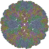
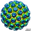
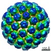
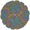
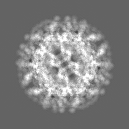
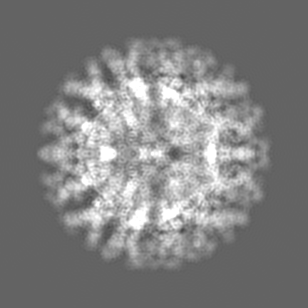
Journal: Sci Adv / Year: 2021 Title: The structure of an infectious immature flavivirus redefines viral architecture and maturation. Authors: Natalee D Newton / Joshua M Hardy / Naphak Modhiran / Leon E Hugo / Alberto A Amarilla / Summa Bibby / Hariprasad Venugopal / Jessica J Harrison / Renee J Traves / Roy A Hall / Jody Hobson- ...Authors: Natalee D Newton / Joshua M Hardy / Naphak Modhiran / Leon E Hugo / Alberto A Amarilla / Summa Bibby / Hariprasad Venugopal / Jessica J Harrison / Renee J Traves / Roy A Hall / Jody Hobson-Peters / Fasséli Coulibaly / Daniel Watterson / Abstract: Flaviviruses are the cause of severe human diseases transmitted by mosquitoes and ticks. These viruses use a potent fusion machinery to enter target cells that needs to be restrained during viral ...Flaviviruses are the cause of severe human diseases transmitted by mosquitoes and ticks. These viruses use a potent fusion machinery to enter target cells that needs to be restrained during viral assembly and egress. A molecular chaperone, premembrane (prM) maintains the virus particles in an immature, fusion-incompetent state until they exit the cell. Taking advantage of an insect virus that produces particles that are both immature and infectious, we determined the structure of the first immature flavivirus with a complete spike by cryo-electron microscopy. Unexpectedly, the prM chaperone forms a supporting pillar that maintains the immature spike in an asymmetric and upright state, primed for large rearrangements upon acidification. The collapse of the spike along a path defined by the prM chaperone is required, and its inhibition by a multivalent immunoglobulin M blocks infection. The revised architecture and collapse model are likely to be conserved across flaviviruses.
History
Deposition
Jan 21, 2021
-
Header (metadata) release
May 26, 2021
-
Map release
May 26, 2021
-
Update
May 26, 2021
-
Current status
May 26, 2021
Processing site: RCSB / Status: Released
-
Structure visualization
Movie
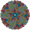
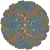
Surface view with section colored by density value
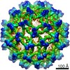
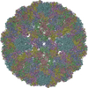
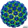
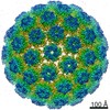
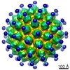
Entire : Binjari virus complexed with pr-specific Fab 2A7
Entire
Name: Binjari virus complexed with pr-specific Fab 2A7
Components
Complex: Binjari virus complexed with pr-specific Fab 2A7
Complex: 2A7 Fab
Virus: Binjari virus
-
Supramolecule #1: Binjari virus complexed with pr-specific Fab 2A7
Supramolecule
Name: Binjari virus complexed with pr-specific Fab 2A7 / type: complex / ID: 1 / Parent: 0 Details: M and E from Binjari virus form a complex that assembles into an asymmetric trimeric spike of prM-E heterodimers. The organization of the immature BinJV particles is T=1 with an asymmetric ...Details: M and E from Binjari virus form a complex that assembles into an asymmetric trimeric spike of prM-E heterodimers. The organization of the immature BinJV particles is T=1 with an asymmetric spike as the basic building block. The 2A7 Fab recognises an epitope on the pr region of BinJV prM and binds the virus with full ocupancy (1:1)
Molecular weight
Theoretical: 22 MDa
-
Supramolecule #3: 2A7 Fab
Supramolecule
Name: 2A7 Fab / type: complex / ID: 3 / Parent: 1 Details: 2A7 Fab fragment generated by cleavage of recombinantly produced IgG 2A7 antibody
Source (natural)
Organism: Mus musculus (house mouse)
Recombinant expression
Organism: Cricetulus griseus (Chinese hamster) / Recombinant cell: CHO
Molecular weight
Theoretical: 50 KDa
-
Supramolecule #2: Binjari virus
Supramolecule
Name: Binjari virus / type: virus / ID: 2 / Parent: 1 Details: M and E from Binjari virus form a complex that assembles into an asymmetric trimeric spike of prM-E heterodimers. The organization of the immature BinJV particles is T=1 with an asymmetric ...Details: M and E from Binjari virus form a complex that assembles into an asymmetric trimeric spike of prM-E heterodimers. The organization of the immature BinJV particles is T=1 with an asymmetric spike as the basic building block. NCBI-ID: 2305258 / Sci species name: Binjari virus / Sci species strain: BFTA20 / Virus type: VIRION / Virus isolate: SPECIES / Virus enveloped: Yes / Virus empty: No
Host (natural)
Organism: Ochlerotatus normanensis (mosquito)
Molecular weight
Theoretical: 22 MDa
-
Experimental details
-
Structure determination
Method
cryo EM
Processing
single particle reconstruction
Aggregation state
particle
-
Sample preparation
Concentration
2 mg/mL
Buffer
pH: 8 Component:
Concentration
Formula
Name
10.0 mM
(HOCH2)3CNH2
Tris
120.0 mM
NaCl
sodium chloride
1.0 mM
C10H16N2O8
EDTA
Grid
Model: PELCO Ultrathin Carbon with Lacey Carbon / Material: COPPER / Mesh: 400 / Support film - Material: CARBON / Support film - topology: HOLEY / Pretreatment - Type: GLOW DISCHARGE
Vitrification
Cryogen name: ETHANE / Chamber humidity: 100 % / Chamber temperature: 277.15 K / Instrument: FEI VITROBOT MARK II
Details
Purified BinJV was complexed with 2A7-Fab at a molar ration of 2:1 Fab:E protein and incubated at 4 C for 2 h.
-
Electron microscopy
Microscope
FEI TECNAI F30
Image recording
Film or detector model: GATAN K2 SUMMIT (4k x 4k) / Detector mode: COUNTING / Digitization - Frames/image: 1-50 / Number grids imaged: 1 / Number real images: 54 / Average exposure time: 5.0 sec. / Average electron dose: 21.95 e/Å2
Electron beam
Acceleration voltage: 300 kV / Electron source: FIELD EMISSION GUN
Electron optics
Calibrated magnification: 50000 / Illumination mode: OTHER / Imaging mode: BRIGHT FIELD / Cs: 2.0 mm
In the structure databanks used in Yorodumi, some data are registered as the other names, "COVID-19 virus" and "2019-nCoV". Here are the details of the virus and the list of structure data.
Jan 31, 2019. EMDB accession codes are about to change! (news from PDBe EMDB page)
EMDB accession codes are about to change! (news from PDBe EMDB page)
The allocation of 4 digits for EMDB accession codes will soon come to an end. Whilst these codes will remain in use, new EMDB accession codes will include an additional digit and will expand incrementally as the available range of codes is exhausted. The current 4-digit format prefixed with “EMD-” (i.e. EMD-XXXX) will advance to a 5-digit format (i.e. EMD-XXXXX), and so on. It is currently estimated that the 4-digit codes will be depleted around Spring 2019, at which point the 5-digit format will come into force.
The EM Navigator/Yorodumi systems omit the EMD- prefix.
Related info.:Q: What is EMD? / ID/Accession-code notation in Yorodumi/EM Navigator
Yorodumi is a browser for structure data from EMDB, PDB, SASBDB, etc.
This page is also the successor to EM Navigator detail page, and also detail information page/front-end page for Omokage search.
The word "yorodu" (or yorozu) is an old Japanese word meaning "ten thousand". "mi" (miru) is to see.
Related info.:EMDB / PDB / SASBDB / Comparison of 3 databanks / Yorodumi Search / Aug 31, 2016. New EM Navigator & Yorodumi / Yorodumi Papers / Jmol/JSmol / Function and homology information / Changes in new EM Navigator and Yorodumi
 Movie
Movie Controller


 Open data
 Basic information
 Map data
 Sample
 Function and homology information
 Binjari virus
 Authors
 Australia, 1 items
  Citation
 Structure visualization
 Downloads & links
 emd_23330.png
 http://ftp.pdbj.org/pub/emdb/structures/EMD-23330
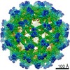


 Z (Sec.)
 Y (Row.)
 X (Col.)


 Sample components
 Cricetulus griseus (Chinese hamster) / Recombinant cell: CHO
 Ochlerotatus normanensis (mosquito)
 Processing
 Electron microscopy
 FIELD EMISSION GUN