Movie
Movie Controller


+ Open data

- Basic information
| Entry | Database: EMDB / ID: EMD-23327 | |||||||||
|---|---|---|---|---|---|---|---|---|---|---|
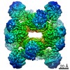
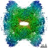
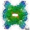
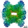
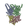
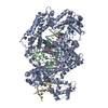
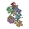
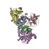
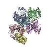
| Title | Cyanophycin synthetase from A. baylyi DSM587 with ATP | |||||||||
 Map data Map data | Cyanophycin synthetase from A. baylyi DSM587 with ATP map | |||||||||
 Sample Sample |
| |||||||||
 Keywords Keywords | Cyanophycin / CphA1 / ATP-grasp / LIGASE | |||||||||
| Function / homology |  Function and homology information Function and homology informationcyanophycin synthase (L-aspartate-adding) / cyanophycin synthase (L-arginine-adding) / cyanophycin synthetase activity (L-aspartate-adding) / cyanophycin synthetase activity (L-arginine-adding) / ATP binding / metal ion binding Similarity search - Function | |||||||||
| Biological species |  Acinetobacter baylyi ADP1 (bacteria) / Acinetobacter baylyi (strain ATCC 33305 / BD413 / ADP1) (bacteria) Acinetobacter baylyi ADP1 (bacteria) / Acinetobacter baylyi (strain ATCC 33305 / BD413 / ADP1) (bacteria) | |||||||||
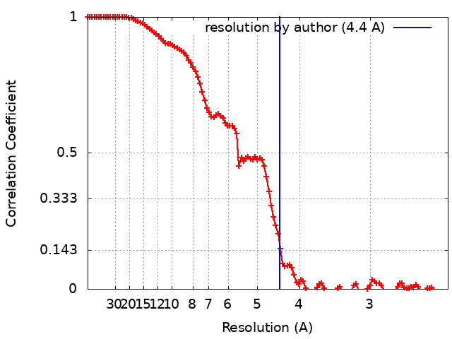
| Method | single particle reconstruction / cryo EM / Resolution: 4.4 Å | |||||||||
 Authors Authors | Sharon I / Haque AS / Lahiri I / Leschziner A / Schmeing TM | |||||||||
 Citation Citation | Journal: Nat Chem Biol / Year: 2021 Title: Structures and function of the amino acid polymerase cyanophycin synthetase. Authors: Itai Sharon / Asfarul S Haque / Marcel Grogg / Indrajit Lahiri / Dieter Seebach / Andres E Leschziner / Donald Hilvert / T Martin Schmeing /    Abstract: Cyanophycin is a natural biopolymer produced by a wide range of bacteria, consisting of a chain of poly-L-Asp residues with L-Arg residues attached to the β-carboxylate sidechains by isopeptide ...Cyanophycin is a natural biopolymer produced by a wide range of bacteria, consisting of a chain of poly-L-Asp residues with L-Arg residues attached to the β-carboxylate sidechains by isopeptide bonds. Cyanophycin is synthesized from ATP, aspartic acid and arginine by a homooligomeric enzyme called cyanophycin synthetase (CphA1). CphA1 has domains that are homologous to glutathione synthetases and muramyl ligases, but no other structural information has been available. Here, we present cryo-electron microscopy and X-ray crystallography structures of cyanophycin synthetases from three different bacteria, including cocomplex structures of CphA1 with ATP and cyanophycin polymer analogs at 2.6 Å resolution. These structures reveal two distinct tetrameric architectures, show the configuration of active sites and polymer-binding regions, indicate dynamic conformational changes and afford insight into catalytic mechanism. Accompanying biochemical interrogation of substrate binding sites, catalytic centers and oligomerization interfaces combine with the structures to provide a holistic understanding of cyanophycin biosynthesis. | |||||||||
| History |
|
- Structure visualization
| Movie |
Movie viewer |
|---|---|
| Structure viewer | EM map: SurfViewMolmilJmol/JSmol |
| Supplemental images |

- Downloads & links
-EMDB archive
| Map data | emd_23327.map.gz | 117.9 MB | EMDB map data format | |
|---|---|---|---|---|
| Header (meta data) | emd-23327-v30.xmlemd-23327.xml | 14.5 KB 14.5 KB | Display Display | EMDB header |
| FSC (resolution estimation) | emd_23327_fsc.xml | 11.1 KB | Display | FSC data file |
| Images | 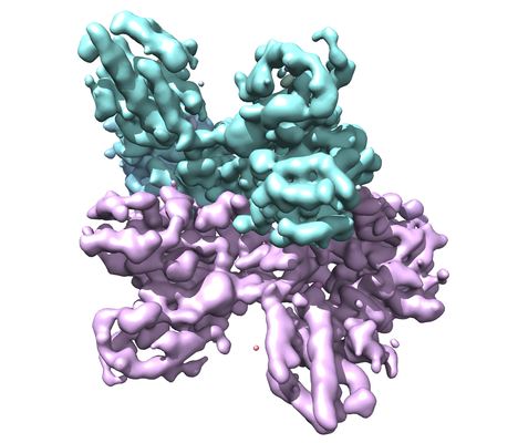 emd_23327.png emd_23327.png | 162.4 KB | ||
| Masks | emd_23327_msk_1.map | 125 MB | Mask map | |
| Filedesc metadata | emd-23327.cif.gz | 5.6 KB | ||
| Others | emd_23327_half_map_1.map.gzemd_23327_half_map_2.map.gz | 116.2 MB 116.2 MB | ||
| Archive directory |  http://ftp.pdbj.org/pub/emdb/structures/EMD-23327ftp://ftp.pdbj.org/pub/emdb/structures/EMD-23327 http://ftp.pdbj.org/pub/emdb/structures/EMD-23327ftp://ftp.pdbj.org/pub/emdb/structures/EMD-23327 | HTTPS FTP |
-Related structure data
| Related structure data |  7lgmMC  7lg5C 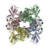 7lgjC 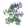 7lgnC 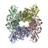 7lgqC M: atomic model generated by this map C: citing same article ( |
|---|---|
| Similar structure data |


-Links
| EMDB pages | EMDB (EBI/PDBe) / EMDataResource |
|---|---|
| Related items in Molecule of the Month |


-Map
| File | Download / File: emd_23327.map.gz / Format: CCP4 / Size: 125 MB / Type: IMAGE STORED AS FLOATING POINT NUMBER (4 BYTES) | ||||||||||||||||||||||||||||||||||||||||||||||||||||||||||||||||||||
|---|---|---|---|---|---|---|---|---|---|---|---|---|---|---|---|---|---|---|---|---|---|---|---|---|---|---|---|---|---|---|---|---|---|---|---|---|---|---|---|---|---|---|---|---|---|---|---|---|---|---|---|---|---|---|---|---|---|---|---|---|---|---|---|---|---|---|---|---|---|
| Annotation | Cyanophycin synthetase from A. baylyi DSM587 with ATP map | ||||||||||||||||||||||||||||||||||||||||||||||||||||||||||||||||||||
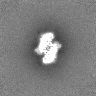
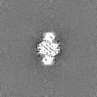
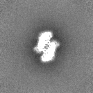
| Projections & slices | Image control
Images are generated by Spider. | ||||||||||||||||||||||||||||||||||||||||||||||||||||||||||||||||||||
| Voxel size | X=Y=Z: 1.16 Å | ||||||||||||||||||||||||||||||||||||||||||||||||||||||||||||||||||||
| Density |
| ||||||||||||||||||||||||||||||||||||||||||||||||||||||||||||||||||||
| Symmetry | Space group: 1 | ||||||||||||||||||||||||||||||||||||||||||||||||||||||||||||||||||||
| Details | EMDB XML:
CCP4 map header:
| ||||||||||||||||||||||||||||||||||||||||||||||||||||||||||||||||||||
 Z (Sec.)
 Y (Row.)
 X (Col.)


-Supplemental data
-Mask #1
| File | emd_23327_msk_1.map | ||||||||||||
|---|---|---|---|---|---|---|---|---|---|---|---|---|---|
| Projections & Slices |
| ||||||||||||
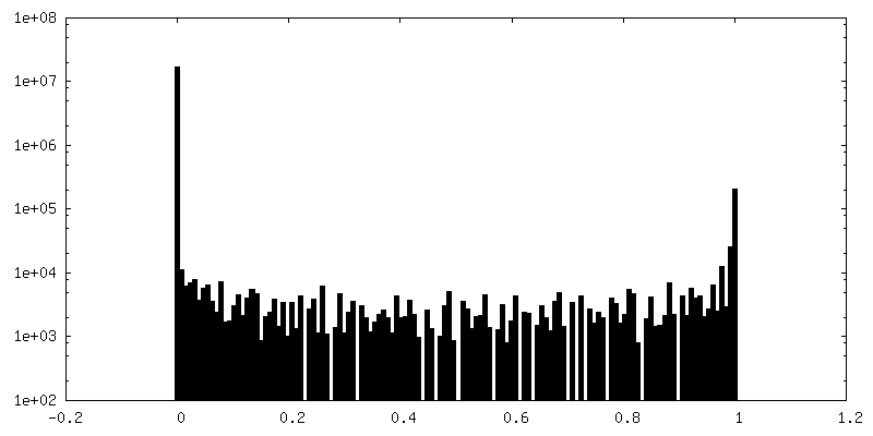
| Density Histograms |


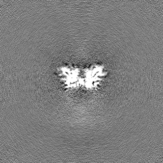
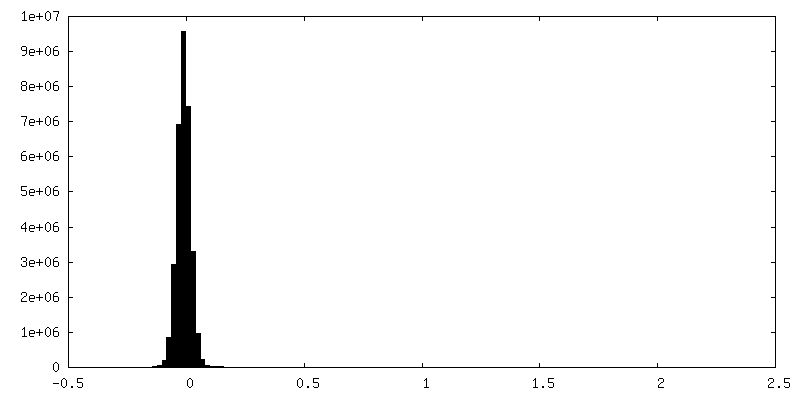
-Half map: Cyanophycin synthetase from A. baylyi DSM587 with ATP half map A
| File | emd_23327_half_map_1.map | ||||||||||||
|---|---|---|---|---|---|---|---|---|---|---|---|---|---|
| Annotation | Cyanophycin synthetase from A. baylyi DSM587 with ATP half map A | ||||||||||||
| Projections & Slices |
| ||||||||||||
| Density Histograms |


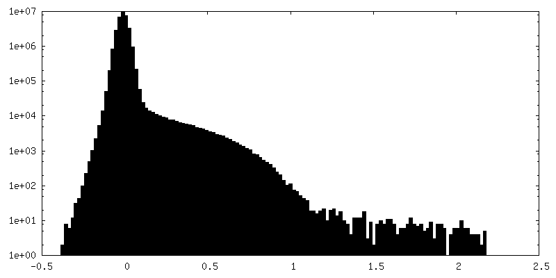
-Half map: Cyanophycin synthetase from A. baylyi DSM587 with ATP half map B
| File | emd_23327_half_map_2.map | ||||||||||||
|---|---|---|---|---|---|---|---|---|---|---|---|---|---|
| Annotation | Cyanophycin synthetase from A. baylyi DSM587 with ATP half map B | ||||||||||||
| Projections & Slices |
| ||||||||||||
| Density Histograms |


- Sample components
-Entire : Cyanophycin synthetase 1 from A. baylyi with ATP
| Entire | Name: Cyanophycin synthetase 1 from A. baylyi with ATP |
|---|---|
| Components |
|
-Supramolecule #1: Cyanophycin synthetase 1 from A. baylyi with ATP
| Supramolecule | Name: Cyanophycin synthetase 1 from A. baylyi with ATP / type: complex / ID: 1 / Parent: 0 / Macromolecule list: #1 |
|---|---|
| Source (natural) | Organism: Acinetobacter baylyi ADP1 (bacteria) |
-Macromolecule #1: Cyanophycin synthase
| Macromolecule | Name: Cyanophycin synthase / type: protein_or_peptide / ID: 1 / Number of copies: 2 / Enantiomer: LEVO / EC number: cyanophycin synthase (L-aspartate-adding) |
|---|---|
| Source (natural) | Organism: Acinetobacter baylyi (strain ATCC 33305 / BD413 / ADP1) (bacteria) Strain: ATCC 33305 / BD413 / ADP1 |
| Molecular weight | Theoretical: 101.783664 KDa |
| Recombinant expression | Organism: |
| Sequence | String: MNIISTSVYV GPNVYASIPL IRLVIDLNPH YITQLASMGS EVLENLEKVI PTLKTEQDAK LQHKLEELRQ APQQQIGELV AILALHLQR LAGQKGGAAF SAYCHEDETE ILYSYESEEI GIEAGEVVCD MLVALAKAHE AGDQIDLNRD VKGFLRYADR F ALGPSALA ...String: MNIISTSVYV GPNVYASIPL IRLVIDLNPH YITQLASMGS EVLENLEKVI PTLKTEQDAK LQHKLEELRQ APQQQIGELV AILALHLQR LAGQKGGAAF SAYCHEDETE ILYSYESEEI GIEAGEVVCD MLVALAKAHE AGDQIDLNRD VKGFLRYADR F ALGPSALA LVQAAEERNI PWYRLNDASL IQVGQGKYQK RIEAALTSGT SHIAVEIAGD KNVCNQLLQD LGLPVPKQRV VY DIDDAVR AARRVGFPVV LKPLDGNHGR GVSVNLTTDE AVEAAFDIAM SEGSAVIVES MLYGDDHRLL VVNGELVAAA RRV PGHIVG DGKHNVEALI EIVNQDPRRG VGHENMLTKI ELDEQALKLL AEKGYDKDSI PAKDEVVYLR RTANISTGGT AIDV TDTIH PENKLMAERA IRAVGLDIGA VDFLTTDITK SYRDIGGGIC EVNAGPGLRM HISPSEGPSR DVGGKIMDML FPQGS QSRV PIAAITGTNG KTTCSRMLAH ILKMAGHVVG QTSTDAVYID GNVTVKGDMT GPVSAKMVLR DPSVDIAVLE TARGGI VRS GLGYQFCDVG AVLNVSSDHL GLGGVDTLDG LAEVKRVIAE VTKDTVVLNA DNAYTLKMAG HSPAKHIMYV TRDAENK LV REHIRLGKRA VVLEKGLNGD QIVIYENGTQ IPLIWTHLIP ATLEGKAIHN VENAMFAAGM AYALGKNLDQ IRIGLRTF D NTFFQSPGRM NVFDKHGFRV ILDYGHNEAA VGAMTELVDR LNPRGRRLLG VTCPGDRRDE DVVAIAAKVA GHFDEYYCH RDDDLRGRAP DETPKIMRDA LIQLGVPESR IHIVEQEEDS LAAVLTEAQV DDLVLFFCEN ITRSWKQIVH FTPEFNIEND HETLELKIA EQGFDIPEGY HAVSNDRGVM ILPRGENLYF QGHHHHHHHH UniProtKB: Cyanophycin synthetase |
-Macromolecule #2: ADENOSINE-5'-TRIPHOSPHATE
| Macromolecule | Name: ADENOSINE-5'-TRIPHOSPHATE / type: ligand / ID: 2 / Number of copies: 2 / Formula: ATP |
|---|---|
| Molecular weight | Theoretical: 507.181 Da |
| Chemical component information |  ChemComp-ATP: |
-Experimental details
-Structure determination
| Method | cryo EM |
|---|---|
 Processing Processing | single particle reconstruction |
| Aggregation state | particle |
-Sample preparation
| Buffer | pH: 8 |
|---|---|
| Vitrification | Cryogen name: ETHANE |
- Electron microscopy
| Microscope | FEI TALOS ARCTICA |
|---|---|
| Image recording | Film or detector model: GATAN K2 SUMMIT (4k x 4k) / Detector mode: SUPER-RESOLUTION / Average electron dose: 57.0 e/Å2 |
| Electron beam | Acceleration voltage: 200 kV / Electron source:  FIELD EMISSION GUN FIELD EMISSION GUN |
| Electron optics | Illumination mode: OTHER / Imaging mode: OTHER |
| Experimental equipment |  Model: Talos Arctica / Image courtesy: FEI Company |