 Movie
Movie Controller


+ Open data

- Basic information
| Entry | Database: EMDB / ID: EMD-23249 | |||||||||||||||
|---|---|---|---|---|---|---|---|---|---|---|---|---|---|---|---|---|
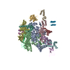
| Title | Cryo-EM structure of PCV2 Replicase bound to ssDNA | |||||||||||||||
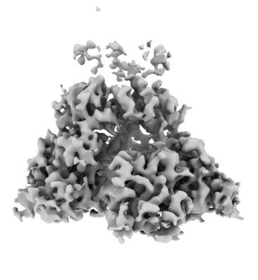
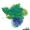
 Map data Map data | Cryo-EM map of PCV2 Replicase at 3.8 Angstrom resolution | |||||||||||||||
 Sample Sample |
| |||||||||||||||
 Keywords Keywords | Rolling circle replication / CRESS-DNA virus / SF3 helicase / Porcine Circovirus / PCV2 / REPLICATION / HYDROLASE-DNA complex | |||||||||||||||
| Function / homology |  Function and homology information Function and homology informationnucleotidyltransferase activity / endonuclease activity / DNA replication / RNA helicase activity / hydrolase activity / host cell nucleus / DNA binding / RNA binding / ATP binding / metal ion binding Similarity search - Function | |||||||||||||||
| Biological species |  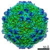 Porcine circovirus 2 / unidentified (others) Porcine circovirus 2 / unidentified (others) | |||||||||||||||
| Method | single particle reconstruction / cryo EM / Resolution: 3.8 Å | |||||||||||||||
 Authors Authors | Khayat R | |||||||||||||||
| Funding support |  United States, 4 items United States, 4 items
| |||||||||||||||
 Citation Citation | Journal: mBio / Year: 2021 Title: Mechanism of DNA Interaction and Translocation by the Replicase of a Circular Rep-Encoding Single-Stranded DNA Virus. Authors: Elvira Tarasova / Sonali Dhindwal / Matthew Popp / Sakeenah Hussain / Reza Khayat / Abstract: Circular Rep-encoding single-stranded DNA (CRESS-DNA) viruses infect members from all three domains of life (, , and ). The replicase (Rep) from these viruses is responsible for initiating rolling ...Circular Rep-encoding single-stranded DNA (CRESS-DNA) viruses infect members from all three domains of life (, , and ). The replicase (Rep) from these viruses is responsible for initiating rolling circle replication (RCR) of their genomes. Rep is a multifunctional enzyme responsible for nicking and ligating ssDNA and unwinding double-stranded DNA (dsDNA). We report the structure of porcine circovirus 2 (PCV2) Rep bound to ADP and single-stranded DNA (ssDNA), and Rep bound to ADP and double-stranded DNA (dsDNA). The structures demonstrate Rep to be a member of the superfamily 3 (SF3) of ATPases Associated with diverse cellular Activities (AAA) superfamily clade 4. At the Rep N terminus is an endonuclease domain () that is responsible for ssDNA nicking and ligation, in the center of Rep is an oligomerization domain () responsible for hexamerization, and at the C terminus is an ATPase domain () responsible for ssDNA/dsDNA interaction and translocation. The Rep binds to DNA such that the faces the replication fork. The six spiral around the DNA to interact with the backbone phosphates from four consecutive nucleotides. Three of the six are able to sense the backbone phosphates from the second strand of dsDNA. Heterogeneous classification of the data demonstrates the and to be mobile. Furthermore, we demonstrate that Rep exhibits basal nucleoside triphosphatase (NTPase) activity. CRESS-DNA viruses encompass a significant portion of the biosphere's virome. However, little is known about the structure of Rep responsible for initiating the RCR of CRESS-DNA viruses. We use cryo-electron microscopy (cryo-EM) to determine the structure of PCV2 Rep in complex with ADP and ss/dsDNA. Our structures demonstrate CRESS-DNA Reps to be SF3 members (clade 4) of the AAA+ superfamily. The structures further provide the mechanism by which CRESS-DNA virus Reps recognize DNA and translocate DNA for genome replication. Our structures also demonstrate the and of PCV2 Rep to be highly mobile. We propose the mobile nature of these domains to be necessary for proper functioning of Reps. We further demonstrate that Reps exhibit basal NTPase activity. Our studies also provide initial insight into the mechanism of RCR. | |||||||||||||||
| History |
|
- Structure visualization
| Movie |
Movie viewer |
|---|---|
| Structure viewer | EM map: SurfViewMolmilJmol/JSmol |
| Supplemental images |
- Downloads & links
-EMDB archive
| Map data | emd_23249.map.gz | 32.8 MB | EMDB map data format | |
|---|---|---|---|---|
| Header (meta data) | emd-23249-v30.xmlemd-23249.xml | 14.6 KB 14.6 KB | Display Display | EMDB header |
| Images |  emd_23249.png emd_23249.png | 53.7 KB | ||
| Filedesc metadata | emd-23249.cif.gz | 6 KB | ||
| Archive directory |  http://ftp.pdbj.org/pub/emdb/structures/EMD-23249ftp://ftp.pdbj.org/pub/emdb/structures/EMD-23249 http://ftp.pdbj.org/pub/emdb/structures/EMD-23249ftp://ftp.pdbj.org/pub/emdb/structures/EMD-23249 | HTTPS FTP |
-Related structure data
| Related structure data |  7larMC  7lasC C: citing same article ( M: atomic model generated by this map |
|---|---|
| Similar structure data |


-Links
| EMDB pages | EMDB (EBI/PDBe) / EMDataResource |
|---|---|
| Related items in Molecule of the Month |

-Map
| File | Download / File: emd_23249.map.gz / Format: CCP4 / Size: 64 MB / Type: IMAGE STORED AS FLOATING POINT NUMBER (4 BYTES) | ||||||||||||||||||||||||||||||||||||||||||||||||||||||||||||
|---|---|---|---|---|---|---|---|---|---|---|---|---|---|---|---|---|---|---|---|---|---|---|---|---|---|---|---|---|---|---|---|---|---|---|---|---|---|---|---|---|---|---|---|---|---|---|---|---|---|---|---|---|---|---|---|---|---|---|---|---|---|
| Annotation | Cryo-EM map of PCV2 Replicase at 3.8 Angstrom resolution | ||||||||||||||||||||||||||||||||||||||||||||||||||||||||||||
| Projections & slices | Image control
Images are generated by Spider. | ||||||||||||||||||||||||||||||||||||||||||||||||||||||||||||
| Voxel size | X=Y=Z: 0.832 Å | ||||||||||||||||||||||||||||||||||||||||||||||||||||||||||||
| Density |
| ||||||||||||||||||||||||||||||||||||||||||||||||||||||||||||
| Symmetry | Space group: 1 | ||||||||||||||||||||||||||||||||||||||||||||||||||||||||||||
| Details | EMDB XML:
CCP4 map header:
| ||||||||||||||||||||||||||||||||||||||||||||||||||||||||||||
 Z (Sec.)
 Y (Row.)
 X (Col.)


-Supplemental data
- Sample components
-Entire : PCV2 Replicase in complex with ssDNA
| Entire | Name: PCV2 Replicase in complex with ssDNA |
|---|---|
| Components |
|
-Supramolecule #1: PCV2 Replicase in complex with ssDNA
| Supramolecule | Name: PCV2 Replicase in complex with ssDNA / type: complex / ID: 1 / Parent: 0 / Macromolecule list: #1-#2 |
|---|---|
| Source (natural) | Organism: Porcine circovirus 2 |
| Molecular weight | Theoretical: 216 KDa |
-Macromolecule #1: ATP-dependent helicase Rep
| Macromolecule | Name: ATP-dependent helicase Rep / type: protein_or_peptide / ID: 1 / Number of copies: 6 / Enantiomer: LEVO EC number: Hydrolases; Acting on acid anhydrides; Acting on acid anhydrides to facilitate cellular and subcellular movement |
|---|---|
| Source (natural) | Organism: Porcine circovirus 2 |
| Molecular weight | Theoretical: 35.8765 KDa |
| Recombinant expression | Organism:  |
| Sequence | String: MPSKKNGRSG PQPHKRWVFT LNNPSEDERK KIRELPISLF DYFIVGEEGN EEGRTPHLQG FANFVKKQTF NKVKWYFGAR CHIEKAKGT DQQNKEYCSK EGNLLMECGA PRSQGQRSDL STAVSTLLES GSLVTVAEQH PVTFVRNFRG LAELLKVSGK M QKRDWKTN ...String: MPSKKNGRSG PQPHKRWVFT LNNPSEDERK KIRELPISLF DYFIVGEEGN EEGRTPHLQG FANFVKKQTF NKVKWYFGAR CHIEKAKGT DQQNKEYCSK EGNLLMECGA PRSQGQRSDL STAVSTLLES GSLVTVAEQH PVTFVRNFRG LAELLKVSGK M QKRDWKTN VHVIVGPPGC GKSKWAANFA DPETTYWKPP RNKWWDGYHG EEVVVIDDFY GWLPWDDLLR LCDRYPLTVE TK GGTVPFL ARSILITSNQ TPLEWYSSTA VPAVEALYRR ITSLVFWKNA TEQSTEEGGQ FVTLSPPCPE FPYEINY UniProtKB: Replication-associated protein |
-Macromolecule #2: DNA (5'-D(P*TP*TP*TP*TP*TP*T)-3')
| Macromolecule | Name: DNA (5'-D(P*TP*TP*TP*TP*TP*T)-3') / type: dna / ID: 2 / Number of copies: 1 / Classification: DNA |
|---|---|
| Source (natural) | Organism: unidentified (others) |
| Molecular weight | Theoretical: 1.780199 KDa |
| Sequence | String: (DT)(DT)(DT)(DT)(DT)(DT) |
-Macromolecule #3: ADENOSINE-5'-DIPHOSPHATE
| Macromolecule | Name: ADENOSINE-5'-DIPHOSPHATE / type: ligand / ID: 3 / Number of copies: 4 / Formula: ADP |
|---|---|
| Molecular weight | Theoretical: 427.201 Da |
| Chemical component information |  ChemComp-ADP: |
-Macromolecule #4: MAGNESIUM ION
| Macromolecule | Name: MAGNESIUM ION / type: ligand / ID: 4 / Number of copies: 4 / Formula: MG |
|---|---|
| Molecular weight | Theoretical: 24.305 Da |
-Experimental details
-Structure determination
| Method | cryo EM |
|---|---|
 Processing Processing | single particle reconstruction |
| Aggregation state | particle |
-Sample preparation
| Buffer | pH: 8.2 Details: 20 mM HEPES, pH 8.2, 500 mM NaCl, 0.2 mM TCEP, 10 mM MgCl2 |
|---|---|
| Grid | Model: UltrAuFoil / Material: GOLD / Mesh: 400 / Support film - Material: GOLD / Support film - topology: HOLEY / Pretreatment - Type: PLASMA CLEANING / Pretreatment - Time: 30 sec. |
| Vitrification | Cryogen name: ETHANE / Chamber humidity: 100 % / Chamber temperature: 277 K / Instrument: FEI VITROBOT MARK IV Details: 4 uL samples were applied to the grids for 3 seconds and blotted using a blot force of 2, a blot time of 4 seconds, and a drain time of 0 seconds.. |
| Details | Sample was monodisperse according to size exclusion chromatography |
- Electron microscopy
| Microscope | FEI TITAN KRIOS |
|---|---|
| Image recording | Film or detector model: GATAN K2 SUMMIT (4k x 4k) / Detector mode: COUNTING / Number grids imaged: 1 / Average exposure time: 6.0 sec. / Average electron dose: 71.8 e/Å2 |
| Electron beam | Acceleration voltage: 300 kV / Electron source:  FIELD EMISSION GUN FIELD EMISSION GUN |
| Electron optics | Illumination mode: SPOT SCAN / Imaging mode: BRIGHT FIELD |
| Experimental equipment |  Model: Titan Krios / Image courtesy: FEI Company |
+Image processing
-Atomic model buiding 1
| Refinement | Space: REAL / Protocol: FLEXIBLE FIT / Overall B value: 65.3 / Target criteria: Correlation coefficient |
|---|---|
| Output model | PDB-7lar: |
