ムービー
ムービー コントローラー


+ データを開く

- 基本情報
基本情報
| 登録情報 | データベース: EMDB / ID: EMD-2318 | |||||||||
|---|---|---|---|---|---|---|---|---|---|---|
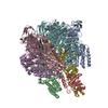
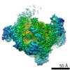
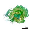
| タイトル | Single particle cryo-microscopy reconstruction of the isolated Manduca sexta V1 domain | |||||||||
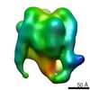
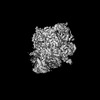
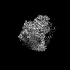
 マップデータ マップデータ | V1 domain without the C subunit bound | |||||||||
 試料 試料 |
| |||||||||
 キーワード キーワード | V-ATPase / atp synthase / membrane complex | |||||||||
| 生物種 |  Manduca sexta (蝶・蛾) Manduca sexta (蝶・蛾) | |||||||||
| 手法 | 単粒子再構成法 / クライオ電子顕微鏡法 / ネガティブ染色法 / 解像度: 20.0 Å | |||||||||
 データ登録者 データ登録者 | Muench SP / Scheres SHW / Huss M / Phillips C / Vitavska O / Wieczorek H / Trinick J / Harrison MA | |||||||||
 引用 引用 | ジャーナル: J Mol Biol / 年: 2014 タイトル: Subunit positioning and stator filament stiffness in regulation and power transmission in the V1 motor of the Manduca sexta V-ATPase. 著者: Stephen P Muench / Sjors H W Scheres / Markus Huss / Clair Phillips / Olga Vitavska / Helmut Wieczorek / John Trinick / Michael A Harrison /   要旨: The vacuolar H(+)-ATPase (V-ATPase) is an ATP-driven proton pump essential to the function of eukaryotic cells. Its cytoplasmic V1 domain is an ATPase, normally coupled to membrane-bound proton pump ...The vacuolar H(+)-ATPase (V-ATPase) is an ATP-driven proton pump essential to the function of eukaryotic cells. Its cytoplasmic V1 domain is an ATPase, normally coupled to membrane-bound proton pump Vo via a rotary mechanism. How these asymmetric motors are coupled remains poorly understood. Low energy status can trigger release of V1 from the membrane and curtail ATP hydrolysis. To investigate the molecular basis for these processes, we have carried out cryo-electron microscopy three-dimensional reconstruction of deactivated V1 from Manduca sexta. In the resulting model, three peripheral stalks that are parts of the mechanical stator of the V-ATPase are clearly resolved as unsupported filaments in the same conformations as in the holoenzyme. They are likely therefore to have inherent stiffness consistent with a role as flexible rods in buffering elastic power transmission between the domains of the V-ATPase. Inactivated V1 adopted a homogeneous resting state with one open active site adjacent to the stator filament normally linked to the H subunit. Although present at 1:1 stoichiometry with V1, both recombinant subunit C reconstituted with V1 and its endogenous subunit H were poorly resolved in three-dimensional reconstructions, suggesting structural heterogeneity in the region at the base of V1 that could indicate positional variability. If the position of H can vary, existing mechanistic models of deactivation in which it binds to and locks the axle of the V-ATPase rotary motor would need to be re-evaluated. | |||||||||
| 履歴 |
|
- 構造の表示
構造の表示
| ムービー |
 ムービービューア ムービービューア |
|---|---|
| 構造ビューア | EMマップ: SurfViewMolmilJmol/JSmol |
| 添付画像 |

- ダウンロードとリンク
-EMDBアーカイブ
| マップデータ | emd_2318.map.gz | 461.2 KB | EMDBマップデータ形式 | |
|---|---|---|---|---|
| ヘッダ (付随情報) | emd-2318-v30.xmlemd-2318.xml | 11.6 KB 11.6 KB | 表示 表示 | EMDBヘッダ |
| 画像 |  emd_2318.png emd_2318.png | 112.6 KB | ||
| アーカイブディレクトリ |  http://ftp.pdbj.org/pub/emdb/structures/EMD-2318ftp://ftp.pdbj.org/pub/emdb/structures/EMD-2318 http://ftp.pdbj.org/pub/emdb/structures/EMD-2318ftp://ftp.pdbj.org/pub/emdb/structures/EMD-2318 | HTTPS FTP |
-検証レポート
| 文書・要旨 | emd_2318_validation.pdf.gz | 200.7 KB | 表示 | EMDB検証レポート |
|---|---|---|---|---|
| 文書・詳細版 | emd_2318_full_validation.pdf.gz | 199.8 KB | 表示 | |
| XML形式データ | emd_2318_validation.xml.gz | 5.1 KB | 表示 | |
| アーカイブディレクトリ | https://ftp.pdbj.org/pub/emdb/validation_reports/EMD-2318ftp://ftp.pdbj.org/pub/emdb/validation_reports/EMD-2318 | HTTPS FTP |
-関連構造データ

-リンク
| EMDBのページ | EMDB (EBI/PDBe) / EMDataResource |
|---|
-マップ
| ファイル | ダウンロード / ファイル: emd_2318.map.gz / 形式: CCP4 / 大きさ: 825.2 KB / タイプ: IMAGE STORED AS FLOATING POINT NUMBER (4 BYTES) | ||||||||||||||||||||||||||||||||||||||||||||||||||||||||||||||||||||
|---|---|---|---|---|---|---|---|---|---|---|---|---|---|---|---|---|---|---|---|---|---|---|---|---|---|---|---|---|---|---|---|---|---|---|---|---|---|---|---|---|---|---|---|---|---|---|---|---|---|---|---|---|---|---|---|---|---|---|---|---|---|---|---|---|---|---|---|---|---|
| 注釈 | V1 domain without the C subunit bound | ||||||||||||||||||||||||||||||||||||||||||||||||||||||||||||||||||||
| 投影像・断面図 | 画像のコントロール
画像は Spider により作成 | ||||||||||||||||||||||||||||||||||||||||||||||||||||||||||||||||||||
| ボクセルのサイズ | X=Y=Z: 4.36 Å | ||||||||||||||||||||||||||||||||||||||||||||||||||||||||||||||||||||
| 密度 |
| ||||||||||||||||||||||||||||||||||||||||||||||||||||||||||||||||||||
| 対称性 | 空間群: 1 | ||||||||||||||||||||||||||||||||||||||||||||||||||||||||||||||||||||
| 詳細 | EMDB XML:
CCP4マップ ヘッダ情報:
| ||||||||||||||||||||||||||||||||||||||||||||||||||||||||||||||||||||
 Z (Sec.)
 Y (Row.)
 X (Col.)


-添付データ
- 試料の構成要素
-全体 : M. sexta isolated V1 domain without subunit C
| 全体 | 名称: M. sexta isolated V1 domain without subunit C |
|---|---|
| 要素 |
|
-超分子 #1000: M. sexta isolated V1 domain without subunit C
| 超分子 | 名称: M. sexta isolated V1 domain without subunit C / タイプ: sample / ID: 1000 詳細: The sample was monodisperse and not freeze thawed to maintain V1 integrity 集合状態: One V1 bound to a monomer of subunit C / Number unique components: 1 |
|---|---|
| 分子量 | 実験値: 650 KDa / 理論値: 650 KDa / 手法: Mass Spec |
-分子 #1: Vacuolar ATPase isolated V1 domain
| 分子 | 名称: Vacuolar ATPase isolated V1 domain / タイプ: protein_or_peptide / ID: 1 / Name.synonym: V1 / 詳細: No C subunit / コピー数: 1 / 集合状態: 3A,B,E,G subunits and 1 D,F,H subunit / 組換発現: No |
|---|---|
| 由来(天然) | 生物種: Manduca sexta (蝶・蛾) / 別称: Tobacco Hornworm / 組織: midgut epithelium |
| 分子量 | 実験値: 650 KDa / 理論値: 650 KDa |
-実験情報
-構造解析
| 手法 | ネガティブ染色法, クライオ電子顕微鏡法 |
|---|---|
 解析 解析 | 単粒子再構成法 |
| 試料の集合状態 | particle |
-試料調製
| 濃度 | 1 mg/mL |
|---|---|
| 緩衝液 | pH: 8.1 / 詳細: 150mM NaCl, 20mM Tris-HCL, 0.01% C12E10 |
| 染色 | タイプ: NEGATIVE 詳細: negative stain data was collected using 1% w/v uranyl acetate for 1 minute. |
| グリッド | 詳細: UV glow discharged grids, 400 mesh |
| 凍結 | 凍結剤: ETHANE / チャンバー内湿度: 100 % / チャンバー内温度: 120 K / 装置: FEI VITROBOT MARK IV 手法: Grids were blotted for 6 seconds with a 6 second drain time |
- 電子顕微鏡法
| 顕微鏡 | FEI TECNAI F20 |
|---|---|
| 温度 | 最低: 90 K / 最高: 110 K / 平均: 100 K |
| アライメント法 | Legacy - 非点収差: Astigmatism corrected for each image in focus mode at 100,000 times magnification |
| 詳細 | FEI Low dose mode |
| 日付 | 2011年4月14日 |
| 撮影 | カテゴリ: CCD フィルム・検出器のモデル: GENERIC GATAN (4k x 4k) 平均電子線量: 16 e/Å2 / 詳細: All collected on Gatan 4K x 4K CCD |
| 電子線 | 加速電圧: 200 kV / 電子線源:  FIELD EMISSION GUN FIELD EMISSION GUN |
| 電子光学系 | 倍率(補正後): 69000 / 照射モード: FLOOD BEAM / 撮影モード: BRIGHT FIELD / Cs: 2.0 mm / 最大 デフォーカス(公称値): 0.0035 µm / 最小 デフォーカス(公称値): 0.001 µm / 倍率(公称値): 50000 |
| 試料ステージ | 試料ホルダー: Nitrogen cooled 3350 holder / 試料ホルダーモデル: GATAN LIQUID NITROGEN |
| 実験機器 |  モデル: Tecnai F20 / 画像提供: FEI Company |
-画像解析
| 詳細 | The particles were handpicked in BOXER |
|---|---|
| CTF補正 | 詳細: Relion |
| 最終 再構成 | 想定した対称性 - 点群: C1 (非対称) / アルゴリズム: OTHER / 解像度のタイプ: BY AUTHOR / 解像度: 20.0 Å / 解像度の算出法: OTHER / ソフトウェア - 名称: Relion 詳細: Maximum likelihood in Relion using the MLF3D protocol 使用した粒子像数: 16500 |
| 最終 角度割当 | 詳細: Relion angular sampling 10 degrees |
-原子モデル構築 1
| 初期モデル | PDB ID: Chain - #0 - Chain ID: A / Chain - #1 - Chain ID: B / Chain - #2 - Chain ID: C / Chain - #3 - Chain ID: D / Chain - #4 - Chain ID: E / Chain - #5 - Chain ID: F / Chain - #6 - Chain ID: G / Chain - #7 - Chain ID: H |
|---|---|
| ソフトウェア | 名称: Chimera |
| 詳細 | The domains were fitted using Chimera |
| 精密化 | 空間: REAL / プロトコル: RIGID BODY FIT / 当てはまり具合の基準: best fit |