 ムービー
ムービー コントローラー


+ データを開く

- 基本情報
基本情報
| 登録情報 | データベース: EMDB / ID: EMD-22982 | |||||||||
|---|---|---|---|---|---|---|---|---|---|---|
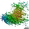
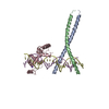
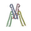
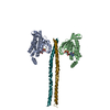
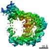
| タイトル | Nucleocapsid protein from the SARS-CoV-2 coronavirus | |||||||||
 マップデータ マップデータ | Nucleocapsid protein from the SARS-CoV-2 Coronavirus | |||||||||
 試料 試料 | SARS-CoV-2 != Severe acute respiratory syndrome coronavirus 2 SARS-CoV-2
| |||||||||
| 生物種 |   Severe acute respiratory syndrome coronavirus 2 (ウイルス) Severe acute respiratory syndrome coronavirus 2 (ウイルス) | |||||||||
| 手法 | 単粒子再構成法 / クライオ電子顕微鏡法 / 解像度: 4.3 Å | |||||||||
 データ登録者 データ登録者 | Casasanta MA / Kelly DF | |||||||||
 引用 引用 | ジャーナル: Nanoscale / 年: 2021 タイトル: Microchip-based structure determination of low-molecular weight proteins using cryo-electron microscopy. 著者: Michael A Casasanta / G M Jonaid / Liam Kaylor / William Y Luqiu / Maria J Solares / Mariah L Schroen / William J Dearnaley / Jarad Wilson / Madeline J Dukes / Deborah F Kelly /  要旨: Interest in cryo-Electron Microscopy (EM) imaging has skyrocketed in recent years due to its pristine views of macromolecules and materials. As advances in instrumentation and computing algorithms ...Interest in cryo-Electron Microscopy (EM) imaging has skyrocketed in recent years due to its pristine views of macromolecules and materials. As advances in instrumentation and computing algorithms spurred this progress, there is renewed focus to address specimen-related challenges. Here we contribute a microchip-based toolkit to perform complementary structural and biochemical analysis on low-molecular weight proteins. As a model system, we used the SARS-CoV-2 nucleocapsid (N) protein (48 kDa) due to its stability and important role in therapeutic development. Cryo-EM structures of the N protein monomer revealed a flexible N-terminal "top hat" motif and a helical-rich C-terminal domain. To complement our structural findings, we engineered microchip-based immunoprecipitation assays that led to the discovery of the first antibody binding site on the N protein. The data also facilitated molecular modeling of a variety of pandemic and common cold-related coronavirus proteins. Such insights may guide future pandemic-preparedness protocols through immuno-engineering strategies to mitigate viral outbreaks. | |||||||||
| 履歴 |
|
- 構造の表示
構造の表示
| ムービー |
 ムービービューア ムービービューア |
|---|---|
| 構造ビューア | EMマップ: SurfViewMolmilJmol/JSmol |
| 添付画像 |

- ダウンロードとリンク
-EMDBアーカイブ
| マップデータ | emd_22982.map.gz | 304.2 KB | EMDBマップデータ形式 | |
|---|---|---|---|---|
| ヘッダ (付随情報) | emd-22982-v30.xmlemd-22982.xml | 12.6 KB 12.6 KB | 表示 表示 | EMDBヘッダ |
| 画像 | 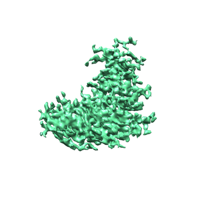 emd_22982.png emd_22982.png | 79.6 KB | ||
| アーカイブディレクトリ |  http://ftp.pdbj.org/pub/emdb/structures/EMD-22982ftp://ftp.pdbj.org/pub/emdb/structures/EMD-22982 http://ftp.pdbj.org/pub/emdb/structures/EMD-22982ftp://ftp.pdbj.org/pub/emdb/structures/EMD-22982 | HTTPS FTP |
-検証レポート
| 文書・要旨 | emd_22982_validation.pdf.gz | 306.7 KB | 表示 | EMDB検証レポート |
|---|---|---|---|---|
| 文書・詳細版 | emd_22982_full_validation.pdf.gz | 306.2 KB | 表示 | |
| XML形式データ | emd_22982_validation.xml.gz | 4.9 KB | 表示 | |
| アーカイブディレクトリ | https://ftp.pdbj.org/pub/emdb/validation_reports/EMD-22982ftp://ftp.pdbj.org/pub/emdb/validation_reports/EMD-22982 | HTTPS FTP |
-関連構造データ


-リンク
| EMDBのページ | EMDB (EBI/PDBe) / EMDataResource |
|---|
-マップ
| ファイル | ダウンロード / ファイル: emd_22982.map.gz / 形式: CCP4 / 大きさ: 12.9 MB / タイプ: IMAGE STORED AS FLOATING POINT NUMBER (4 BYTES) | ||||||||||||||||||||||||||||||||||||||||||||||||||||||||||||
|---|---|---|---|---|---|---|---|---|---|---|---|---|---|---|---|---|---|---|---|---|---|---|---|---|---|---|---|---|---|---|---|---|---|---|---|---|---|---|---|---|---|---|---|---|---|---|---|---|---|---|---|---|---|---|---|---|---|---|---|---|---|
| 注釈 | Nucleocapsid protein from the SARS-CoV-2 Coronavirus | ||||||||||||||||||||||||||||||||||||||||||||||||||||||||||||
| ボクセルのサイズ | X=Y=Z: 0.93 Å | ||||||||||||||||||||||||||||||||||||||||||||||||||||||||||||
| 密度 |
| ||||||||||||||||||||||||||||||||||||||||||||||||||||||||||||
| 対称性 | 空間群: 1 | ||||||||||||||||||||||||||||||||||||||||||||||||||||||||||||
| 詳細 | EMDB XML:
CCP4マップ ヘッダ情報:
| ||||||||||||||||||||||||||||||||||||||||||||||||||||||||||||
-添付データ
- 試料の構成要素
-全体 : SARS-CoV-2
| 全体 | 名称: SARS-CoV-2 (ウイルス) |
|---|---|
| 要素 |
|
-超分子 #1: Severe acute respiratory syndrome coronavirus 2
| 超分子 | 名称: Severe acute respiratory syndrome coronavirus 2 / タイプ: virus / ID: 1 / 親要素: 0 / NCBI-ID: 2697049 / 生物種: Severe acute respiratory syndrome coronavirus 2 / ウイルスタイプ: VIRION / ウイルス・単離状態: STRAIN / ウイルス・エンベロープ: Yes / ウイルス・中空状態: Yes |
|---|---|
| 宿主 | 生物種:  Homo sapiens (ヒト) Homo sapiens (ヒト) |
| Host system | 生物種:  |
| 分子量 | 実験値: 50 KDa |
-実験情報
-構造解析
| 手法 | クライオ電子顕微鏡法 |
|---|---|
 解析 解析 | 単粒子再構成法 |
| 試料の集合状態 | particle |
-試料調製
| 濃度 | 0.1 mg/mL | |||||||||||||||
|---|---|---|---|---|---|---|---|---|---|---|---|---|---|---|---|---|
| 緩衝液 | pH: 7.5 構成要素:
詳細: Solutions were made fresh before diluting protein and adding to the EM grids | |||||||||||||||
| グリッド | モデル: Homemade / 材質: SILICON NITRIDE / 支持フィルム - トポロジー: CONTINUOUS / 支持フィルム - Film thickness: 20.0 nm | |||||||||||||||
| 凍結 | 凍結剤: ETHANE / チャンバー内湿度: 95 % / チャンバー内温度: 293 K / 装置: FEI VITROBOT MARK III / 詳細: blot for 7.5 seconds before plunging. | |||||||||||||||
| 詳細 | Sample was monodisperse on a silicon nitride microchip coated with 25% Ni-NTA lipids. |
- 電子顕微鏡法
| 顕微鏡 | TFS TALOS F200C |
|---|---|
| 温度 | 最低: 75.0 K / 最高: 83.0 K |
| 撮影 | フィルム・検出器のモデル: DIRECT ELECTRON DE-12 (4k x 3k) 検出モード: INTEGRATING / 撮影したグリッド数: 3 / 平均露光時間: 0.25 sec. / 平均電子線量: 5.0 e/Å2 詳細: Images were collected in movie mode with 0.25 second exposures at 30 frames per second. |
| 電子線 | 加速電圧: 200 kV / 電子線源:  FIELD EMISSION GUN FIELD EMISSION GUN |
| 電子光学系 | 照射モード: FLOOD BEAM / 撮影モード: BRIGHT FIELD / Cs: 2.7 mm / 最大 デフォーカス(公称値): 1.5 µm / 最小 デフォーカス(公称値): 0.5 µm / 倍率(公称値): 45000 |
| 試料ステージ | 試料ホルダーモデル: GATAN 626 SINGLE TILT LIQUID NITROGEN CRYO TRANSFER HOLDER ホルダー冷却材: NITROGEN |
| 実験機器 |  モデル: Tecnai F20 / 画像提供: FEI Company |
+画像解析
-原子モデル構築 1
| 精密化 | 空間: REAL / プロトコル: FLEXIBLE FIT |
|---|
