 Movie
Movie Controller


+ Open data

- Basic information
| Entry | Database: EMDB / ID: EMD-22727 | |||||||||
|---|---|---|---|---|---|---|---|---|---|---|
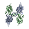
| Title | EsxE-EsxF pentameric pore | |||||||||
 Map data Map data | EsxE/EsxF (Mycobacterium tuberculosis) hetero-pentameric pore produced recombinantly in E.coli. Single particle reconstruction in 1% uranyl formate. Reconstruction performed with EMAN2.2. | |||||||||
 Sample Sample |
| |||||||||
| Biological species |   Mycobacterium tuberculosis (bacteria) / Mycobacterium tuberculosis H37RV (bacteria) Mycobacterium tuberculosis (bacteria) / Mycobacterium tuberculosis H37RV (bacteria) | |||||||||
| Method | single particle reconstruction / negative staining / Resolution: 17.6 Å | |||||||||
 Authors Authors | Tak U / Dokland T / Niederweis M | |||||||||
| Funding support |  United States, 1 items United States, 1 items
| |||||||||
 Citation Citation | Journal: Nat Commun / Year: 2021 Title: Pore-forming Esx proteins mediate toxin secretion by Mycobacterium tuberculosis. Authors: Uday Tak / Terje Dokland / Michael Niederweis / Abstract: Mycobacterium tuberculosis secretes the tuberculosis necrotizing toxin (TNT) to kill host cells. Here, we show that the WXG100 proteins EsxE and EsxF are essential for TNT secretion. EsxE and EsxF ...Mycobacterium tuberculosis secretes the tuberculosis necrotizing toxin (TNT) to kill host cells. Here, we show that the WXG100 proteins EsxE and EsxF are essential for TNT secretion. EsxE and EsxF form a water-soluble heterodimer (EsxEF) that assembles into oligomers and long filaments, binds to membranes, and forms stable membrane-spanning channels. Electron microscopy of EsxEF reveals mainly pentameric structures with a central pore. Mutations of both WXG motifs and of a GXW motif do not affect dimerization, but abolish pore formation, membrane deformation and TNT secretion. The WXG/GXW mutants are locked in conformations with altered thermostability and solvent exposure, indicating that the WXG/GXW motifs are molecular switches controlling membrane interaction and pore formation. EsxF is accessible on the bacterial cell surface, suggesting that EsxEF form an outer membrane channel for toxin export. Thus, our study reveals a protein secretion mechanism in bacteria that relies on pore formation by small WXG proteins. | |||||||||
| History |
|
- Structure visualization
| Movie |
 Movie viewer Movie viewer |
|---|---|
| Structure viewer | EM map: SurfViewMolmilJmol/JSmol |
| Supplemental images |

- Downloads & links
-EMDB archive
| Map data | emd_22727.map.gz | 12.3 MB | EMDB map data format | |
|---|---|---|---|---|
| Header (meta data) | emd-22727-v30.xmlemd-22727.xml | 15.5 KB 15.5 KB | Display Display | EMDB header |
| FSC (resolution estimation) | emd_22727_fsc.xml | 6.9 KB | Display | FSC data file |
| Images |  emd_22727.png emd_22727.png | 59.2 KB | ||
| Masks | emd_22727_msk_1.mapemd_22727_msk_2.map | 12.9 MB 12.9 MB | Mask map | |
| Others | emd_22727_half_map_1.map.gzemd_22727_half_map_2.map.gz | 12.4 MB 12.4 MB | ||
| Archive directory |  http://ftp.pdbj.org/pub/emdb/structures/EMD-22727ftp://ftp.pdbj.org/pub/emdb/structures/EMD-22727 http://ftp.pdbj.org/pub/emdb/structures/EMD-22727ftp://ftp.pdbj.org/pub/emdb/structures/EMD-22727 | HTTPS FTP |
-Related structure data
| Similar structure data |
|---|

-Links
| EMDB pages | EMDB (EBI/PDBe) / EMDataResource |
|---|
-Map
| File | Download / File: emd_22727.map.gz / Format: CCP4 / Size: 12.9 MB / Type: IMAGE STORED AS FLOATING POINT NUMBER (4 BYTES) | ||||||||||||||||||||||||||||||||||||||||||||||||||||||||||||||||||||
|---|---|---|---|---|---|---|---|---|---|---|---|---|---|---|---|---|---|---|---|---|---|---|---|---|---|---|---|---|---|---|---|---|---|---|---|---|---|---|---|---|---|---|---|---|---|---|---|---|---|---|---|---|---|---|---|---|---|---|---|---|---|---|---|---|---|---|---|---|---|
| Annotation | EsxE/EsxF (Mycobacterium tuberculosis) hetero-pentameric pore produced recombinantly in E.coli. Single particle reconstruction in 1% uranyl formate. Reconstruction performed with EMAN2.2. | ||||||||||||||||||||||||||||||||||||||||||||||||||||||||||||||||||||
| Projections & slices | Image control
Images are generated by Spider. | ||||||||||||||||||||||||||||||||||||||||||||||||||||||||||||||||||||
| Voxel size | X=Y=Z: 1.23 Å | ||||||||||||||||||||||||||||||||||||||||||||||||||||||||||||||||||||
| Density |
| ||||||||||||||||||||||||||||||||||||||||||||||||||||||||||||||||||||
| Symmetry | Space group: 1 | ||||||||||||||||||||||||||||||||||||||||||||||||||||||||||||||||||||
| Details | EMDB XML:
CCP4 map header:
| ||||||||||||||||||||||||||||||||||||||||||||||||||||||||||||||||||||
 Z (Sec.)
 Y (Row.)
 X (Col.)


-Supplemental data
-Mask #1
| File | emd_22727_msk_1.map | ||||||||||||
|---|---|---|---|---|---|---|---|---|---|---|---|---|---|
| Projections & Slices |
| ||||||||||||
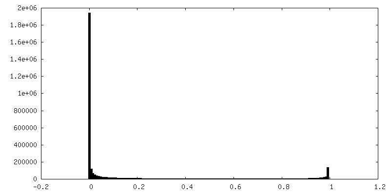
| Density Histograms |


-Mask #2
| File | emd_22727_msk_2.map | ||||||||||||
|---|---|---|---|---|---|---|---|---|---|---|---|---|---|
| Projections & Slices |
| ||||||||||||
| Density Histograms |


-Half map: Half map even
| File | emd_22727_half_map_1.map | ||||||||||||
|---|---|---|---|---|---|---|---|---|---|---|---|---|---|
| Annotation | Half map even | ||||||||||||
| Projections & Slices |
| ||||||||||||
| Density Histograms |


-Half map: Half map odd
| File | emd_22727_half_map_2.map | ||||||||||||
|---|---|---|---|---|---|---|---|---|---|---|---|---|---|
| Annotation | Half map odd | ||||||||||||
| Projections & Slices |
| ||||||||||||
| Density Histograms |


- Sample components
-Entire : Hetero-pentameric pore complex formed from EsxE/EsxF heterodimers...
| Entire | Name: Hetero-pentameric pore complex formed from EsxE/EsxF heterodimers at pH 6.5. |
|---|---|
| Components |
|
-Supramolecule #1: Hetero-pentameric pore complex formed from EsxE/EsxF heterodimers...
| Supramolecule | Name: Hetero-pentameric pore complex formed from EsxE/EsxF heterodimers at pH 6.5. type: complex / ID: 1 / Parent: 0 / Macromolecule list: all |
|---|---|
| Source (natural) | Organism: Mycobacterium tuberculosis (bacteria) / Strain: H37Rv / Location in cell: Membrane, Periplasm, Secreted |
| Recombinant expression | Organism: |
-Macromolecule #1: EsxE/EsxF Pentamer
| Macromolecule | Name: EsxE/EsxF Pentamer / type: other / ID: 1 Details: Hetero-Pentameric pore formed from hetero-oligomers of EsxE/EsxF. Classification: other |
|---|---|
| Source (natural) | Organism: Mycobacterium tuberculosis H37RV (bacteria) |
| Sequence | String: MGADDTLRVE PAVMQGFAAS LDGAAEHLAV QLAELDAQVG QMLGGWRGAS GSAYGSAWEL WHRGAGEVQL GLSMLAAAIA HAGAGYQHNE TASAQVLREV GGG VDPTVL ADAVARMAEF GRHVEELVAE IESLVTRLHV TWTGEGAAAH AEAQRHWAAG EAMMRQALAQ ...String: MGADDTLRVE PAVMQGFAAS LDGAAEHLAV QLAELDAQVG QMLGGWRGAS GSAYGSAWEL WHRGAGEVQL GLSMLAAAIA HAGAGYQHNE TASAQVLREV GGG VDPTVL ADAVARMAEF GRHVEELVAE IESLVTRLHV TWTGEGAAAH AEAQRHWAAG EAMMRQALAQ LTAAGQSAHA NYTGAMATNL GMWS |
| Recombinant expression | Organism: |
-Experimental details
-Structure determination
| Method | negative staining |
|---|---|
 Processing Processing | single particle reconstruction |
| Aggregation state | particle |
-Sample preparation
| Concentration | 0.008 mg/mL |
|---|---|
| Buffer | pH: 6.5 / Component - Formula: NaCl / Component - Name: sodium phosphate Details: Solutions were made fresh, autoclaved, and filter sterilized to prevent microbial contamination. 25 mM sodium phosophate, 150 mM NaCl pH 6.5 |
| Staining | Type: NEGATIVE / Material: Uranyl Formate Details: 3 microliters of sample was placed on glow discharged grids for 1 minute, then blotted with filter paper, washed / blotted 2x with milli-q water, then stained with 1% uranyl formate for 10 ...Details: 3 microliters of sample was placed on glow discharged grids for 1 minute, then blotted with filter paper, washed / blotted 2x with milli-q water, then stained with 1% uranyl formate for 10 seconds, blotted, followed by staining with 1% uranyl formate for 1 minute. The stain was then blotted and the grid was allowed to air dry. |
| Details | This sample was well dispersed. |
- Electron microscopy
| Microscope | FEI TECNAI F20 |
|---|---|
| Image recording | Film or detector model: GATAN K3 (6k x 4k) / Number grids imaged: 1 / Number real images: 52 / Average exposure time: 1.0 sec. / Average electron dose: 30.0 e/Å2 |
| Electron beam | Acceleration voltage: 200 kV / Electron source:  FIELD EMISSION GUN FIELD EMISSION GUN |
| Electron optics | Illumination mode: SPOT SCAN / Imaging mode: BRIGHT FIELD |
| Experimental equipment |  Model: Tecnai F20 / Image courtesy: FEI Company |

