 Movie
Movie Controller


+ Open data

- Basic information
| Entry | Database: EMDB / ID: EMD-22688 | |||||||||
|---|---|---|---|---|---|---|---|---|---|---|
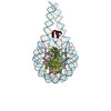
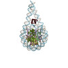
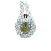
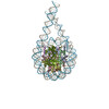
| Title | cryo-EM map of di-chromatosome containing H1.4 | |||||||||
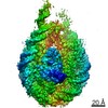
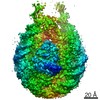
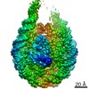
 Map data Map data | cryo-EM map of di-chromatosome containing human linker histone H1.4 | |||||||||
 Sample Sample |
| |||||||||
| Biological species |  human (human) / Homo sapiens (human) / human (human) / Homo sapiens (human) /  Mougeotia scalaris (plant) Mougeotia scalaris (plant) | |||||||||
| Method | single particle reconstruction / cryo EM / Resolution: 9.73 Å | |||||||||
 Authors Authors | Zhou B-R / Bai Y | |||||||||
| Funding support |  United States, 1 items United States, 1 items
| |||||||||
 Citation Citation | Journal: Mol Cell / Year: 2021 Title: Distinct Structures and Dynamics of Chromatosomes with Different Human Linker Histone Isoforms. Authors: Bing-Rui Zhou / Hanqiao Feng / Seyit Kale / Tara Fox / Htet Khant / Natalia de Val / Rodolfo Ghirlando / Anna R Panchenko / Yawen Bai /   Abstract: The repeating structural unit of metazoan chromatin is the chromatosome, a nucleosome bound to a linker histone, H1. There are 11 human H1 isoforms with diverse cellular functions, but how they ...The repeating structural unit of metazoan chromatin is the chromatosome, a nucleosome bound to a linker histone, H1. There are 11 human H1 isoforms with diverse cellular functions, but how they interact with the nucleosome remains elusive. Here, we determined the cryoelectron microscopy (cryo-EM) structures of chromatosomes containing 197 bp DNA and three different human H1 isoforms, respectively. The globular domains of all three H1 isoforms bound to the nucleosome dyad. However, the flanking/linker DNAs displayed substantial distinct dynamic conformations. Nuclear magnetic resonance (NMR) and H1 tail-swapping cryo-EM experiments revealed that the C-terminal tails of the H1 isoforms mainly controlled the flanking DNA orientations. We also observed partial ordering of the core histone H2A C-terminal and H3 N-terminal tails in the chromatosomes. Our results provide insights into the structures and dynamics of the chromatosomes and have implications for the structure and function of chromatin. | |||||||||
| History |
|
- Structure visualization
| Movie |
 Movie viewer Movie viewer |
|---|---|
| Structure viewer | EM map: SurfViewMolmilJmol/JSmol |
| Supplemental images |

- Downloads & links
-EMDB archive
| Map data | emd_22688.map.gz | 4.1 MB | EMDB map data format | |
|---|---|---|---|---|
| Header (meta data) | emd-22688-v30.xmlemd-22688.xml | 15.6 KB 15.6 KB | Display Display | EMDB header |
| Images | 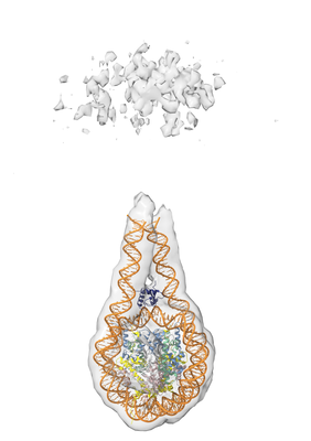 emd_22688.png emd_22688.png | 57.1 KB | ||
| Archive directory |  http://ftp.pdbj.org/pub/emdb/structures/EMD-22688ftp://ftp.pdbj.org/pub/emdb/structures/EMD-22688 http://ftp.pdbj.org/pub/emdb/structures/EMD-22688ftp://ftp.pdbj.org/pub/emdb/structures/EMD-22688 | HTTPS FTP |
-Related structure data


-Links
| EMDB pages | EMDB (EBI/PDBe) / EMDataResource |
|---|
-Map
| File | Download / File: emd_22688.map.gz / Format: CCP4 / Size: 8 MB / Type: IMAGE STORED AS FLOATING POINT NUMBER (4 BYTES) | ||||||||||||||||||||||||||||||||||||||||||||||||||||||||||||||||||||
|---|---|---|---|---|---|---|---|---|---|---|---|---|---|---|---|---|---|---|---|---|---|---|---|---|---|---|---|---|---|---|---|---|---|---|---|---|---|---|---|---|---|---|---|---|---|---|---|---|---|---|---|---|---|---|---|---|---|---|---|---|---|---|---|---|---|---|---|---|---|
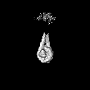
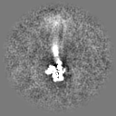
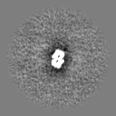
| Annotation | cryo-EM map of di-chromatosome containing human linker histone H1.4 | ||||||||||||||||||||||||||||||||||||||||||||||||||||||||||||||||||||
| Projections & slices | Image control
Images are generated by Spider. | ||||||||||||||||||||||||||||||||||||||||||||||||||||||||||||||||||||
| Voxel size | X=Y=Z: 4.75 Å | ||||||||||||||||||||||||||||||||||||||||||||||||||||||||||||||||||||
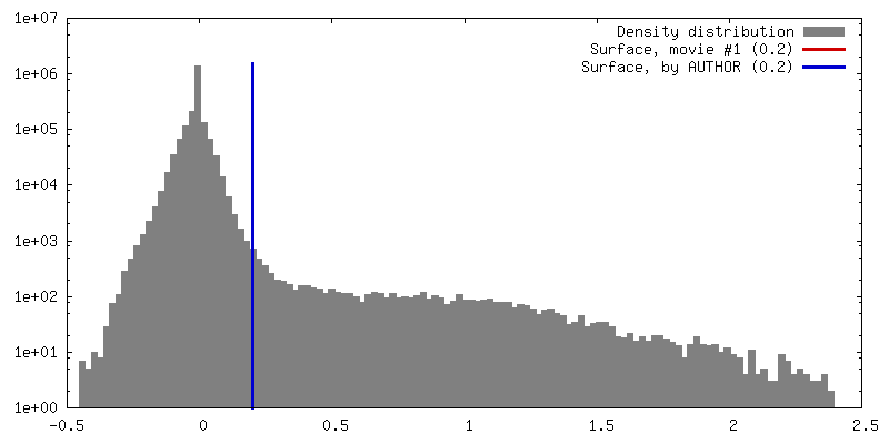
| Density |
| ||||||||||||||||||||||||||||||||||||||||||||||||||||||||||||||||||||
| Symmetry | Space group: 1 | ||||||||||||||||||||||||||||||||||||||||||||||||||||||||||||||||||||
| Details | EMDB XML:
CCP4 map header:
| ||||||||||||||||||||||||||||||||||||||||||||||||||||||||||||||||||||
 Z (Sec.)
 Y (Row.)
 X (Col.)


-Supplemental data
- Sample components
-Entire : di-chromatosome containing human linker histone H1.4
| Entire | Name: di-chromatosome containing human linker histone H1.4 |
|---|---|
| Components |
|
-Supramolecule #1: di-chromatosome containing human linker histone H1.4
| Supramolecule | Name: di-chromatosome containing human linker histone H1.4 / type: complex / ID: 1 / Parent: 0 / Macromolecule list: all |
|---|---|
| Source (natural) | Organism: human (human) |
| Recombinant expression | Organism:  |
-Macromolecule #1: human histone H3.1
| Macromolecule | Name: human histone H3.1 / type: protein_or_peptide / ID: 1 / Enantiomer: LEVO |
|---|---|
| Source (natural) | Organism: Homo sapiens (human) |
| Recombinant expression | Organism: |
| Sequence | String: MARTKQTARK STGGKAPRK Q LATKAARK SA PATGGVK KPH RYRPGT VALR EIRRY QKSTE LLIR KLPFQR LVR EIAQDFK TD LRFQSSAV M ALQEACEAY LVGLFEDTNL CAIHAKRVT I MPKDIQLA RR IRGERA |
-Macromolecule #2: human histone H4
| Macromolecule | Name: human histone H4 / type: protein_or_peptide / ID: 2 / Enantiomer: LEVO |
|---|---|
| Source (natural) | Organism: Homo sapiens (human) |
| Sequence | String: MSGRGKGGKG LGKGGAKRH R KVLRDNIQ GI TKPAIRR LAR RGGVKR ISGL IYEET RGVLK VFLE NVIRDA VTY TEHAKRK TV TAMDVVYA L KRQGRTLYG FGG |
-Macromolecule #3: human histone H2A1
| Macromolecule | Name: human histone H2A1 / type: protein_or_peptide / ID: 3 / Enantiomer: LEVO |
|---|---|
| Source (natural) | Organism: Homo sapiens (human) |
| Recombinant expression | Organism: |
| Sequence | String: MSGRGKQGGK ARAKAKTRS S RAGLQFPV GR VHRLLRK GNY SERVGA GAPV YLAAV LEYLT AEIL ELAGNA ARD NKKTRII PR HLQLAIRN D EELNKLLGR VTIAQGGVLP NIQAVLLPK K TESHHKAK GK |
-Macromolecule #4: human histone H2B1J
| Macromolecule | Name: human histone H2B1J / type: protein_or_peptide / ID: 4 / Enantiomer: LEVO |
|---|---|
| Source (natural) | Organism: Homo sapiens (human) |
| Recombinant expression | Organism: |
| Sequence | String: MPEPAKSAP A PKKGSKKA VT KAQKKDG KKR KRSRKE SYSI YVYKV LKQVH PDTG ISSKAM GIM NSFVNDI FE RIAGEASR L AHYNKRSTI TSREIQTAVR LLLPGELAK H AVSEGTKA VT KYTSAK |
-Macromolecule #5: DNA (394-MER)
| Macromolecule | Name: DNA (394-MER) / type: protein_or_peptide / ID: 5 / Enantiomer: LEVO |
|---|---|
| Source (natural) | Organism: Homo sapiens (human) |
| Recombinant expression | Organism: |
| Sequence | String: GGGCTGGACC CTATACGCGG CCGCCCTGGA GAATCCCGGT GCCGAGGCCG CTCAATTGGT CGTAGACAGC TCTAGCACCG CTTAAACGCA CGTACGCGCT GTCCCCCGCG TTTTAACCGC CAAGGGGATT ACTCCCTAGT CTCCAGGCAC GTGTCAGATA TATACATCCT ...String: GGGCTGGACC CTATACGCGG CCGCCCTGGA GAATCCCGGT GCCGAGGCCG CTCAATTGGT CGTAGACAGC TCTAGCACCG CTTAAACGCA CGTACGCGCT GTCCCCCGCG TTTTAACCGC CAAGGGGATT ACTCCCTAGT CTCCAGGCAC GTGTCAGATA TATACATCCT GTGCATGTAT TGAACAGCGA CCACAGTACT CTGGACCCTA TACGCGGCCG CCCTGGAGAA TCCCGGTGCC GAGGCCGCTC AATTGGTCGT AGACAGCTCT AGCACCGCTT AAACGCACGT ACGCGCTGTC CCCCGCGTTT TAACCGCCAA GGGGATTACT CCCTAGTCTC CAGGCACGTG TCAGATATAT ACATCCTGTG CATGTATTGA ACAGCGACCA CCCC |
-Macromolecule #6: DNA(394-MER)
| Macromolecule | Name: DNA(394-MER) / type: protein_or_peptide / ID: 6 / Enantiomer: LEVO |
|---|---|
| Source (natural) | Organism: Homo sapiens (human) |
| Recombinant expression | Organism: |
| Sequence | String: GGGGTGGTCG CTGTTCAATA CATGCACAGG ATGTATATAT CTGACACGTG CCTGGAGACT AGGGAGTAAT CCCCTTGGCG GTTAAAACGC GGGGGACAGC GCGTACGTGC GTTTAAGCGG TGCTAGAGCT GTCTACGACC AATTGAGCGG CCTCGGCACC GGGATTCTCC ...String: GGGGTGGTCG CTGTTCAATA CATGCACAGG ATGTATATAT CTGACACGTG CCTGGAGACT AGGGAGTAAT CCCCTTGGCG GTTAAAACGC GGGGGACAGC GCGTACGTGC GTTTAAGCGG TGCTAGAGCT GTCTACGACC AATTGAGCGG CCTCGGCACC GGGATTCTCC AGGGCGGCCG CGTATAGGGT CCAGAGTACT GTGGTCGCTG TTCAATACAT GCACAGGATG TATATATCTG ACACGTGCCT GGAGACTAGG GAGTAATCCC CTTGGCGGTT AAAACGCGGG GGACAGCGCG TACGTGCGTT TAAGCGGTGC TAGAGCTGTC TACGACCAAT TGAGCGGCCT CGGCACCGGG ATTCTCCAGG GCGGCCGCGT ATAGGGTCCA GCCC |
-Macromolecule #7: scFv20
| Macromolecule | Name: scFv20 / type: protein_or_peptide / ID: 7 / Enantiomer: LEVO |
|---|---|
| Source (natural) | Organism: Mougeotia scalaris (plant) |
| Recombinant expression | Organism: |
| Sequence | String: MKSSHHHHHH ENLYFQSNAM DIKMTQSPSS MHASLGERVT ITCKASQDIR SYLSWYQQKP WKSPKTLIYY ATSLADGVPS RFSGSGSGQD FSLTINNLES DDTATYYCLQ HGESPYTFGS GTKLEIKRAG GGGSGGGGSG GGGSGGGGSM EVQLQQSGPE LVEPGTSVKM ...String: MKSSHHHHHH ENLYFQSNAM DIKMTQSPSS MHASLGERVT ITCKASQDIR SYLSWYQQKP WKSPKTLIYY ATSLADGVPS RFSGSGSGQD FSLTINNLES DDTATYYCLQ HGESPYTFGS GTKLEIKRAG GGGSGGGGSG GGGSGGGGSM EVQLQQSGPE LVEPGTSVKM PCKASGYTFT SYTIQWVKQT PRQGLEWIGY IYPYNAGTKY NEKFKGKATL TSDKSSSTVY MELSSLTSED SAVYYCARKS SRLRSTLDYW GQGTSVTVSS |
-Macromolecule #8: human histone H1.4
| Macromolecule | Name: human histone H1.4 / type: protein_or_peptide / ID: 8 / Enantiomer: LEVO |
|---|---|
| Source (natural) | Organism: Homo sapiens (human) |
| Recombinant expression | Organism: |
| Sequence | String: MSETAPAAPA APAPAEKTP V KKKARKSA GA AKRKASG PPV SELITK AVAA SKERS GVSLA ALKK ALAAAG YDV EKNNSRI KL GLKSLVSK G TLVQTKGTG ASGSFKLNKK AASGEAKPK A KKAGAAKA KK PAGAAKK PKK ATGAAT PKKS AKKTP ...String: MSETAPAAPA APAPAEKTP V KKKARKSA GA AKRKASG PPV SELITK AVAA SKERS GVSLA ALKK ALAAAG YDV EKNNSRI KL GLKSLVSK G TLVQTKGTG ASGSFKLNKK AASGEAKPK A KKAGAAKA KK PAGAAKK PKK ATGAAT PKKS AKKTP KKAKK PAAA AGAKKA KSP KKAKAAK PK KAPKSPAK A KAVKPKAAK PKTAKPKAAK PKKAAAKKK |
-Experimental details
-Structure determination
| Method | cryo EM |
|---|---|
 Processing Processing | single particle reconstruction |
| Aggregation state | particle |
-Sample preparation
| Buffer | pH: 7.4 |
|---|---|
| Vitrification | Cryogen name: ETHANE |
- Electron microscopy
| Microscope | FEI TITAN KRIOS |
|---|---|
| Image recording | Film or detector model: GATAN K2 SUMMIT (4k x 4k) / Average electron dose: 40.0 e/Å2 |
| Electron beam | Acceleration voltage: 300 kV / Electron source:  FIELD EMISSION GUN FIELD EMISSION GUN |
| Electron optics | Illumination mode: OTHER / Imaging mode: OTHER |
| Experimental equipment |  Model: Titan Krios / Image courtesy: FEI Company |
-Image processing
| Final reconstruction | Resolution.type: BY AUTHOR / Resolution: 9.73 Å / Resolution method: FSC 0.143 CUT-OFF / Software - Name: cryoSPARC (ver. 2) / Number images used: 10876 |
|---|---|
| Initial angle assignment | Type: NOT APPLICABLE |
| Final angle assignment | Type: NOT APPLICABLE |
