Movie
Movie Controller

[English] 日本語
 Yorodumi
- EMDB-22655: Structure of full-length influenza HA with a head-binding antibod... -
+ Open data

- Basic information
| Entry | Database: EMDB / ID: EMD-22655 | |||||||||
|---|---|---|---|---|---|---|---|---|---|---|
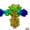
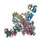
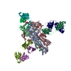
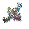
| Title | Structure of full-length influenza HA with a head-binding antibody at pH 5.2, conformation C, central helices splay | |||||||||
 Map data Map data | ||||||||||
 Sample Sample |
| |||||||||
 Keywords Keywords | influenza / virus / hemagglutinin / antibody / low pH / VIRAL PROTEIN | |||||||||
| Function / homology |  Function and homology information Function and homology informationviral budding from plasma membrane / clathrin-dependent endocytosis of virus by host cell / host cell surface receptor binding / fusion of virus membrane with host plasma membrane / fusion of virus membrane with host endosome membrane / viral envelope / virion attachment to host cell / host cell plasma membrane / virion membrane / membrane Similarity search - Function | |||||||||
| Biological species |  Influenza A virus (A/Hong Kong/1/1968(H3N2)) / Influenza A virus (strain A/Hong Kong/1/1968 H3N2) / Influenza A virus (A/Hong Kong/1/1968(H3N2)) / Influenza A virus (strain A/Hong Kong/1/1968 H3N2) /  Homo sapiens (human) Homo sapiens (human) | |||||||||
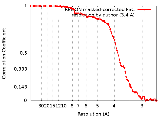
| Method | single particle reconstruction / cryo EM / Resolution: 3.4 Å | |||||||||
 Authors Authors | Gui M / Gao J / Xiang Y | |||||||||
 Citation Citation | Journal: PLoS Pathog / Year: 2020 Title: Structural intermediates in the low pH-induced transition of influenza hemagglutinin. Authors: Jingjing Gao / Miao Gui / Ye Xiang /  Abstract: The hemagglutinin (HA) glycoproteins of influenza viruses play a key role in binding host cell receptors and in mediating virus-host cell membrane fusion during virus infection. Upon virus entry, HA ...The hemagglutinin (HA) glycoproteins of influenza viruses play a key role in binding host cell receptors and in mediating virus-host cell membrane fusion during virus infection. Upon virus entry, HA is triggered by low pH and undergoes large structural rearrangements from a prefusion state to a postfusion state. While structures of prefusion state and postfusion state of HA have been reported, the intermediate structures remain elusive. Here, we report two distinct low pH intermediate conformations of the influenza virus HA using cryo-electron microscopy (cryo-EM). Our results show that a decrease in pH from 7.8 to 5.2 triggers the release of fusion peptides from the binding pockets and then causes a dramatic conformational change in the central helices, in which the membrane-proximal ends of the central helices unwind to an extended form. Accompanying the conformational changes of the central helices, the stem region of the HA undergoes an anticlockwise rotation of 9.5 degrees and a shift of 15 Å. The HA head, after being stabilized by an antibody, remains unchanged compared to the neutral pH state. Thus, the conformational change of the HA stem region observed in our research is likely to be independent of the HA head. These results provide new insights into the structural transition of HA during virus entry. | |||||||||
| History |
|
- Structure visualization
| Movie |
Movie viewer |
|---|---|
| Structure viewer | EM map: SurfViewMolmilJmol/JSmol |
| Supplemental images |

- Downloads & links
-EMDB archive
| Map data | emd_22655.map.gz | 7.3 MB | EMDB map data format | |
|---|---|---|---|---|
| Header (meta data) | emd-22655-v30.xmlemd-22655.xml | 15.2 KB 15.2 KB | Display Display | EMDB header |
| FSC (resolution estimation) | emd_22655_fsc.xml | 10.6 KB | Display | FSC data file |
| Images | 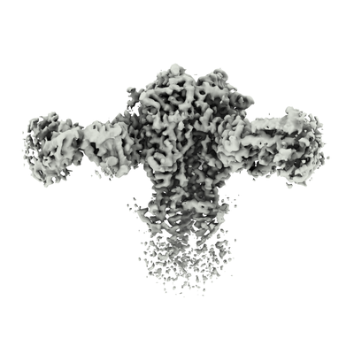 emd_22655.png emd_22655.png | 97 KB | ||
| Masks | emd_22655_msk_1.map | 103 MB | Mask map | |
| Filedesc metadata | emd-22655.cif.gz | 6.1 KB | ||
| Archive directory |  http://ftp.pdbj.org/pub/emdb/structures/EMD-22655ftp://ftp.pdbj.org/pub/emdb/structures/EMD-22655 http://ftp.pdbj.org/pub/emdb/structures/EMD-22655ftp://ftp.pdbj.org/pub/emdb/structures/EMD-22655 | HTTPS FTP |
-Related structure data
| Related structure data |  7k3bMC  7k37C  7k39C  7k3aC C: citing same article ( M: atomic model generated by this map |
|---|---|
| Similar structure data |


-Links
| EMDB pages | EMDB (EBI/PDBe) / EMDataResource |
|---|---|
| Related items in Molecule of the Month |


-Map
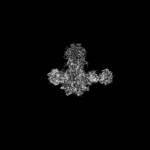
| File | Download / File: emd_22655.map.gz / Format: CCP4 / Size: 103 MB / Type: IMAGE STORED AS FLOATING POINT NUMBER (4 BYTES) | ||||||||||||||||||||||||||||||||||||||||||||||||||||||||||||
|---|---|---|---|---|---|---|---|---|---|---|---|---|---|---|---|---|---|---|---|---|---|---|---|---|---|---|---|---|---|---|---|---|---|---|---|---|---|---|---|---|---|---|---|---|---|---|---|---|---|---|---|---|---|---|---|---|---|---|---|---|---|
| Projections & slices | Image control
Images are generated by Spider. | ||||||||||||||||||||||||||||||||||||||||||||||||||||||||||||
| Voxel size | X=Y=Z: 1.33 Å | ||||||||||||||||||||||||||||||||||||||||||||||||||||||||||||
| Density |
| ||||||||||||||||||||||||||||||||||||||||||||||||||||||||||||
| Symmetry | Space group: 1 | ||||||||||||||||||||||||||||||||||||||||||||||||||||||||||||
| Details | EMDB XML:
CCP4 map header:
| ||||||||||||||||||||||||||||||||||||||||||||||||||||||||||||
 Z (Sec.)
 Y (Row.)
 X (Col.)


-Supplemental data
-Mask #1
| File | emd_22655_msk_1.map | ||||||||||||
|---|---|---|---|---|---|---|---|---|---|---|---|---|---|
| Projections & Slices |
| ||||||||||||
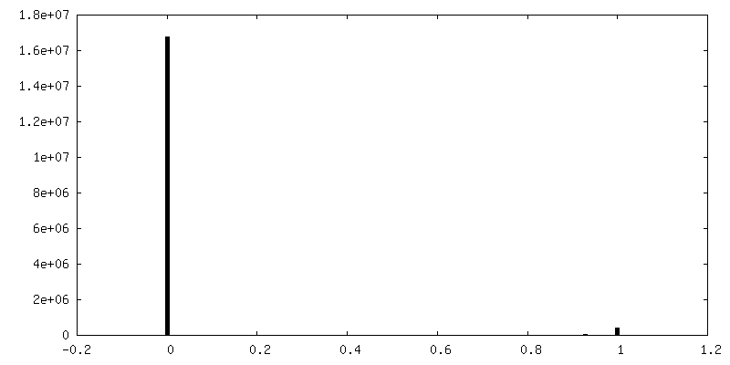
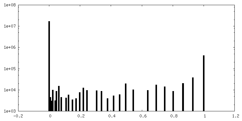
| Density Histograms |


- Sample components
-Entire : complex of hemagglutinin with antibody
| Entire | Name: complex of hemagglutinin with antibody |
|---|---|
| Components |
|
-Supramolecule #1: complex of hemagglutinin with antibody
| Supramolecule | Name: complex of hemagglutinin with antibody / type: complex / ID: 1 / Parent: 0 / Macromolecule list: #1-#4 |
|---|---|
| Source (natural) | Organism: Influenza A virus (A/Hong Kong/1/1968(H3N2)) |
-Supramolecule #2: hemagglutinin
| Supramolecule | Name: hemagglutinin / type: complex / ID: 2 / Parent: 1 / Macromolecule list: #1-#2 |
|---|
-Supramolecule #3: antigen-binding fragment
| Supramolecule | Name: antigen-binding fragment / type: complex / ID: 3 / Parent: 1 / Macromolecule list: #3-#4 |
|---|
-Macromolecule #1: Hemagglutinin
| Macromolecule | Name: Hemagglutinin / type: protein_or_peptide / ID: 1 / Number of copies: 3 / Enantiomer: LEVO |
|---|---|
| Source (natural) | Organism: Influenza A virus (strain A/Hong Kong/1/1968 H3N2) Strain: A/Hong Kong/1/1968 H3N2 |
| Molecular weight | Theoretical: 37.948812 KDa |
| Recombinant expression | Organism: Homo sapiens (human) |
| Sequence | String: MKTIIALSYI FCLALGQDLP GNDNSTATLC LGHHAVPNGT LVKTITDDQI EVTNATELVQ SSSTGKICNN PHRILDGIDC TLIDALLGD PHCDVFQNET WDLFVERSKA FSNCYPYDVP DYASLRSLVA SSGTLEFITE GFTWTGVTQN GGSNACKRGP G SGFFSRLN ...String: MKTIIALSYI FCLALGQDLP GNDNSTATLC LGHHAVPNGT LVKTITDDQI EVTNATELVQ SSSTGKICNN PHRILDGIDC TLIDALLGD PHCDVFQNET WDLFVERSKA FSNCYPYDVP DYASLRSLVA SSGTLEFITE GFTWTGVTQN GGSNACKRGP G SGFFSRLN WLTKSGSTYP VLNVTMPNND NFDKLYIWGV HHPSTNQEQT SLYVQASGRV TVSTRRSQQT IIPNIGSRPW VR GLSSRIS IYWTIVKPGD VLVINSNGNL IAPRGYFKMR TGKSSIMRSD APIDTCISEC ITPNGSIPND KPFQNVNKIT YGA CPKYVK QNTLKLATGM RNVPEKQTR UniProtKB: Hemagglutinin |
-Macromolecule #2: Hemagglutinin
| Macromolecule | Name: Hemagglutinin / type: protein_or_peptide / ID: 2 / Number of copies: 3 / Enantiomer: LEVO |
|---|---|
| Source (natural) | Organism: Influenza A virus (strain A/Hong Kong/1/1968 H3N2) Strain: A/Hong Kong/1/1968 H3N2 |
| Molecular weight | Theoretical: 28.947525 KDa |
| Recombinant expression | Organism: Homo sapiens (human) |
| Sequence | String: GLFGAIAGFI ENGWEGMIDG WYGFRHQNSE GTGQAADLKS TQAAIDQING KLNRVIEKTN EKFHQIEKEF SEVEGRIQDL EKYVEDTKI DLWSYNAELL VALENQHTID LTDSEMNKLF EKTRRQLREN AEDMGNGCFK IYHKCDNACI ESIRNGTYDH D VYRDEALN ...String: GLFGAIAGFI ENGWEGMIDG WYGFRHQNSE GTGQAADLKS TQAAIDQING KLNRVIEKTN EKFHQIEKEF SEVEGRIQDL EKYVEDTKI DLWSYNAELL VALENQHTID LTDSEMNKLF EKTRRQLREN AEDMGNGCFK IYHKCDNACI ESIRNGTYDH D VYRDEALN NRFQIKGVEL KSGYKDWILW ISFAISCFLL CVVLLGFIMW ACQRGNIRCN ICISMGWSHP QFEKGGGARG GS GGGSWSH PQFEKGF UniProtKB: Hemagglutinin |
-Macromolecule #3: antibody Fab light chain
| Macromolecule | Name: antibody Fab light chain / type: protein_or_peptide / ID: 3 / Number of copies: 3 / Enantiomer: LEVO |
|---|---|
| Source (natural) | Organism: Homo sapiens (human) |
| Molecular weight | Theoretical: 23.11241 KDa |
| Recombinant expression | Organism: Homo sapiens (human) |
| Sequence | String: QSVLTQPPSV SGAPGQRVTI SCTGSSSNIG AGYAVHWYQQ LPGTAPKLLI SGNSNRPSGV PDRFSGSKSG TSASLAITGL QAEDEADYY CQSYDSSLSG SVFGGGTKLT VLGRTVAAPS VFIFPPSDEQ LKSGTASVVC LLNNFYPREA KVQWKVDNAL Q SGNSQESV ...String: QSVLTQPPSV SGAPGQRVTI SCTGSSSNIG AGYAVHWYQQ LPGTAPKLLI SGNSNRPSGV PDRFSGSKSG TSASLAITGL QAEDEADYY CQSYDSSLSG SVFGGGTKLT VLGRTVAAPS VFIFPPSDEQ LKSGTASVVC LLNNFYPREA KVQWKVDNAL Q SGNSQESV TEQDSKDSTY SLSSTLTLSK ADYEKHKVYA CEVTHQGLSS PVTKSFNRGE C |
-Macromolecule #4: antibody Fab heavy chain
| Macromolecule | Name: antibody Fab heavy chain / type: protein_or_peptide / ID: 4 / Number of copies: 3 / Enantiomer: LEVO |
|---|---|
| Source (natural) | Organism: Homo sapiens (human) |
| Molecular weight | Theoretical: 25.278605 KDa |
| Recombinant expression | Organism: Homo sapiens (human) |
| Sequence | String: QVQLVQSGAE VKKPGASVTV SCQVSGYTLT SYGLSWVRQA PGQGLEWVGW INTYDGQTKY VKKFQGRVTM TTHTGTNTAY MEMKSLRSD DTAVYYCARV EGVRGVMGFH YYPMDVWGQG TMVTVSSKGP SVFPLAPSSK STSGGTAALG CLVKDYFPEP V TVSWNSGA ...String: QVQLVQSGAE VKKPGASVTV SCQVSGYTLT SYGLSWVRQA PGQGLEWVGW INTYDGQTKY VKKFQGRVTM TTHTGTNTAY MEMKSLRSD DTAVYYCARV EGVRGVMGFH YYPMDVWGQG TMVTVSSKGP SVFPLAPSSK STSGGTAALG CLVKDYFPEP V TVSWNSGA LTSGVHTFPA VLQSSGLYSL SSVVTVPSSS LGTQTYICNV NHKPSNTKVD KKVEPKSCDK GLEVLFQ |
-Macromolecule #8: 2-acetamido-2-deoxy-beta-D-glucopyranose
| Macromolecule | Name: 2-acetamido-2-deoxy-beta-D-glucopyranose / type: ligand / ID: 8 / Number of copies: 6 / Formula: NAG |
|---|---|
| Molecular weight | Theoretical: 221.208 Da |
| Chemical component information |  ChemComp-NAG: |
-Experimental details
-Structure determination
| Method | cryo EM |
|---|---|
 Processing Processing | single particle reconstruction |
| Aggregation state | particle |
-Sample preparation
| Buffer | pH: 5.2 |
|---|---|
| Vitrification | Cryogen name: ETHANE |
- Electron microscopy
| Microscope | FEI TITAN KRIOS |
|---|---|
| Image recording | Film or detector model: GATAN K2 SUMMIT (4k x 4k) / Average electron dose: 50.0 e/Å2 |
| Electron beam | Acceleration voltage: 300 kV / Electron source:  FIELD EMISSION GUN FIELD EMISSION GUN |
| Electron optics | Illumination mode: FLOOD BEAM / Imaging mode: BRIGHT FIELD |
| Experimental equipment |  Model: Titan Krios / Image courtesy: FEI Company |