National Institutes of Health/National Institute of General Medical Sciences
1DP2CA195762-01
United States
Citation
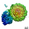
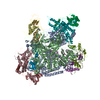
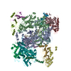
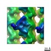
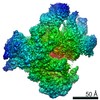
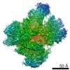
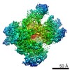
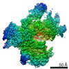
Journal: Cell / Year: 2020 Title: A Structural Model of the Endogenous Human BAF Complex Informs Disease Mechanisms. Authors: Nazar Mashtalir / Hiroshi Suzuki / Daniel P Farrell / Akshay Sankar / Jie Luo / Martin Filipovski / Andrew R D'Avino / Roodolph St Pierre / Alfredo M Valencia / Takashi Onikubo / Robert G ...Authors: Nazar Mashtalir / Hiroshi Suzuki / Daniel P Farrell / Akshay Sankar / Jie Luo / Martin Filipovski / Andrew R D'Avino / Roodolph St Pierre / Alfredo M Valencia / Takashi Onikubo / Robert G Roeder / Yan Han / Yuan He / Jeffrey A Ranish / Frank DiMaio / Thomas Walz / Cigall Kadoch / Abstract: Mammalian SWI/SNF complexes are ATP-dependent chromatin remodeling complexes that regulate genomic architecture. Here, we present a structural model of the endogenously purified human canonical BAF ...Mammalian SWI/SNF complexes are ATP-dependent chromatin remodeling complexes that regulate genomic architecture. Here, we present a structural model of the endogenously purified human canonical BAF complex bound to the nucleosome, generated using cryoelectron microscopy (cryo-EM), cross-linking mass spectrometry, and homology modeling. BAF complexes bilaterally engage the nucleosome H2A/H2B acidic patch regions through the SMARCB1 C-terminal α-helix and the SMARCA4/2 C-terminal SnAc/post-SnAc regions, with disease-associated mutations in either causing attenuated chromatin remodeling activities. Further, we define changes in BAF complex architecture upon nucleosome engagement and compare the structural model of endogenous BAF to those of related SWI/SNF-family complexes. Finally, we assign and experimentally interrogate cancer-associated hot-spot mutations localizing within the endogenous human BAF complex, identifying those that disrupt BAF subunit-subunit and subunit-nucleosome interfaces in the nucleosome-bound conformation. Taken together, this integrative structural approach provides important biophysical foundations for understanding the mechanisms of BAF complex function in normal and disease states.
History
Deposition
Aug 18, 2020
-
Header (metadata) release
Oct 21, 2020
-
Map release
Oct 21, 2020
-
Update
Dec 2, 2020
-
Current status
Dec 2, 2020
Processing site: RCSB / Status: Released
-
Structure visualization
Movie
Surface view with section colored by density value
Support film - #0 - Film type ID: 1 / Support film - #0 - Material: CARBON / Support film - #0 - topology: HOLEY ARRAY / Support film - #1 - Film type ID: 2 / Support film - #1 - Material: CARBON / Support film - #1 - topology: CONTINUOUS / Details: unspecified
Film or detector model: GATAN K2 SUMMIT (4k x 4k) / Detector mode: SUPER-RESOLUTION / Number real images: 14841 / Average exposure time: 15.0 sec. / Average electron dose: 69.0 e/Å2
Electron beam
Acceleration voltage: 200 kV / Electron source: FIELD EMISSION GUN
Electron optics
Illumination mode: FLOOD BEAM / Imaging mode: BRIGHT FIELD
In the structure databanks used in Yorodumi, some data are registered as the other names, "COVID-19 virus" and "2019-nCoV". Here are the details of the virus and the list of structure data.
Jan 31, 2019. EMDB accession codes are about to change! (news from PDBe EMDB page)
EMDB accession codes are about to change! (news from PDBe EMDB page)
The allocation of 4 digits for EMDB accession codes will soon come to an end. Whilst these codes will remain in use, new EMDB accession codes will include an additional digit and will expand incrementally as the available range of codes is exhausted. The current 4-digit format prefixed with “EMD-” (i.e. EMD-XXXX) will advance to a 5-digit format (i.e. EMD-XXXXX), and so on. It is currently estimated that the 4-digit codes will be depleted around Spring 2019, at which point the 5-digit format will come into force.
The EM Navigator/Yorodumi systems omit the EMD- prefix.
Related info.:Q: What is EMD? / ID/Accession-code notation in Yorodumi/EM Navigator
Yorodumi is a browser for structure data from EMDB, PDB, SASBDB, etc.
This page is also the successor to EM Navigator detail page, and also detail information page/front-end page for Omokage search.
The word "yorodu" (or yorozu) is an old Japanese word meaning "ten thousand". "mi" (miru) is to see.
Related info.:EMDB / PDB / SASBDB / Comparison of 3 databanks / Yorodumi Search / Aug 31, 2016. New EM Navigator & Yorodumi / Yorodumi Papers / Jmol/JSmol / Function and homology information / Changes in new EM Navigator and Yorodumi
 Movie
Movie Controller

 Yorodumi
 Open data
 Basic information
 Map data
 Sample
 Homo sapiens (human)
 Authors
 United States, 1 items
  Citation
 Structure visualization
 Movie viewer
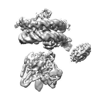
 Downloads & links
 emd_22479.png
 http://ftp.pdbj.org/pub/emdb/structures/EMD-22479

 Z (Sec.)
 Y (Row.)
 X (Col.)


 Sample components
 Processing
 Electron microscopy
 FIELD EMISSION GUN