 ムービー
ムービー コントローラー


+ データを開く

- 基本情報
基本情報
| 登録情報 | データベース: EMDB / ID: EMD-2244 | |||||||||
|---|---|---|---|---|---|---|---|---|---|---|
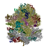
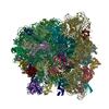
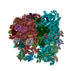
| タイトル | Cryo-electron microscopy of phirsl1 jumbo phage | |||||||||
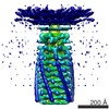
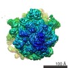
 マップデータ マップデータ | Reconstruction of the helical trunk of phirsl1 tail. A Bfactor of -538 has been applied to the reconstruction as well as a soft-edged mask. | |||||||||
 試料 試料 |
| |||||||||
 キーワード キーワード | Jumbo bacteriophage / phiRSL1 / Ralstonia solanacearum / cryo-electron microscopy / icosahedral capsid / helical tail / T6SS | |||||||||
| 機能・相同性 | Tail sheath protein 機能・相同性情報 機能・相同性情報 | |||||||||
| 生物種 |  Ralstonia phage RSL1 (ファージ) Ralstonia phage RSL1 (ファージ) | |||||||||
| 手法 | らせん対称体再構成法 / クライオ電子顕微鏡法 / ネガティブ染色法 / 解像度: 9.6 Å | |||||||||
 データ登録者 データ登録者 | Effantin G / Hamasaki R / Kawasaki T / Bacia M / Moriscot C / Weissenhorn W / Yamada T / Schoehn G | |||||||||
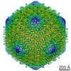
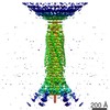
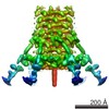
 引用 引用 | ジャーナル: Structure / 年: 2013 タイトル: Cryo-electron microscopy three-dimensional structure of the jumbo phage ΦRSL1 infecting the phytopathogen Ralstonia solanacearum. 著者: Grégory Effantin / Ryosuke Hamasaki / Takeru Kawasaki / Maria Bacia / Christine Moriscot / Winfried Weissenhorn / Takashi Yamada / Guy Schoehn /  要旨: ϕRSL1 jumbo phage belongs to a new class of viruses within the Myoviridae family. Here, we report its three-dimensional structure determined by electron cryo microscopy. The icosahedral capsid, the ...ϕRSL1 jumbo phage belongs to a new class of viruses within the Myoviridae family. Here, we report its three-dimensional structure determined by electron cryo microscopy. The icosahedral capsid, the tail helical portion, and the complete tail appendage were reconstructed separately to resolutions of 9 Å, 9 Å, and 28 Å, respectively. The head is rather complex and formed by at least five different proteins, whereas the major capsid proteins resemble those from HK97, despite low sequence conservation. The helical tail structure demonstrates its close relationship to T4 sheath proteins and provides evidence for an evolutionary link of the inner tail tube to the bacterial type VI secretion apparatus. Long fibers extend from the collar region, and their length is consistent with reaching the host cell surface upon tail contraction. Our structural analyses indicate that ϕRSL1 is an unusual member of the Myoviridae that employs conserved protein machines related to different phages and bacteria. | |||||||||
| 履歴 |
|
- 構造の表示
構造の表示
| ムービー |
ムービービューア |
|---|---|
| 構造ビューア | EMマップ: SurfViewMolmilJmol/JSmol |
| 添付画像 |

- ダウンロードとリンク
-EMDBアーカイブ
| マップデータ | emd_2244.map.gz | 1.8 MB | EMDBマップデータ形式 | |
|---|---|---|---|---|
| ヘッダ (付随情報) | emd-2244-v30.xmlemd-2244.xml | 10.4 KB 10.4 KB | 表示 表示 | EMDBヘッダ |
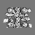
| 画像 | 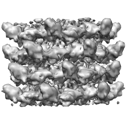 EMD-2244.png EMD-2244.png | 142.3 KB | ||
| アーカイブディレクトリ |  http://ftp.pdbj.org/pub/emdb/structures/EMD-2244ftp://ftp.pdbj.org/pub/emdb/structures/EMD-2244 http://ftp.pdbj.org/pub/emdb/structures/EMD-2244ftp://ftp.pdbj.org/pub/emdb/structures/EMD-2244 | HTTPS FTP |
-検証レポート
| 文書・要旨 | emd_2244_validation.pdf.gz | 251.5 KB | 表示 | EMDB検証レポート |
|---|---|---|---|---|
| 文書・詳細版 | emd_2244_full_validation.pdf.gz | 250.6 KB | 表示 | |
| XML形式データ | emd_2244_validation.xml.gz | 5.2 KB | 表示 | |
| アーカイブディレクトリ | https://ftp.pdbj.org/pub/emdb/validation_reports/EMD-2244ftp://ftp.pdbj.org/pub/emdb/validation_reports/EMD-2244 | HTTPS FTP |
-関連構造データ

-リンク
| EMDBのページ | EMDB (EBI/PDBe) / EMDataResource |
|---|
-マップ
| ファイル | ダウンロード / ファイル: emd_2244.map.gz / 形式: CCP4 / 大きさ: 6.4 MB / タイプ: IMAGE STORED AS FLOATING POINT NUMBER (4 BYTES) | ||||||||||||||||||||||||||||||||||||||||||||||||||||||||||||||||||||
|---|---|---|---|---|---|---|---|---|---|---|---|---|---|---|---|---|---|---|---|---|---|---|---|---|---|---|---|---|---|---|---|---|---|---|---|---|---|---|---|---|---|---|---|---|---|---|---|---|---|---|---|---|---|---|---|---|---|---|---|---|---|---|---|---|---|---|---|---|---|
| 注釈 | Reconstruction of the helical trunk of phirsl1 tail. A Bfactor of -538 has been applied to the reconstruction as well as a soft-edged mask. | ||||||||||||||||||||||||||||||||||||||||||||||||||||||||||||||||||||
| 投影像・断面図 | 画像のコントロール
画像は Spider により作成 | ||||||||||||||||||||||||||||||||||||||||||||||||||||||||||||||||||||
| ボクセルのサイズ | X=Y=Z: 2.26 Å | ||||||||||||||||||||||||||||||||||||||||||||||||||||||||||||||||||||
| 密度 |
| ||||||||||||||||||||||||||||||||||||||||||||||||||||||||||||||||||||
| 対称性 | 空間群: 1 | ||||||||||||||||||||||||||||||||||||||||||||||||||||||||||||||||||||
| 詳細 | EMDB XML:
CCP4マップ ヘッダ情報:
| ||||||||||||||||||||||||||||||||||||||||||||||||||||||||||||||||||||
 Z (Sec.)
 Y (Row.)
 X (Col.)


-添付データ
- 試料の構成要素
-全体 : phirsl1 helical portion of the tail
| 全体 | 名称: phirsl1 helical portion of the tail |
|---|---|
| 要素 |
|
-超分子 #1000: phirsl1 helical portion of the tail
| 超分子 | 名称: phirsl1 helical portion of the tail / タイプ: sample / ID: 1000 / Number unique components: 2 |
|---|
-分子 #1: Tail tube protein
| 分子 | 名称: Tail tube protein / タイプ: protein_or_peptide / ID: 1 / 詳細: UNP B2ZY00 / 組換発現: No / データベース: NCBI |
|---|---|
| 由来(天然) | 生物種: Ralstonia phage RSL1 (ファージ) |
| 分子量 | 理論値: 18 KDa |
-分子 #2: Tail sheath protein
| 分子 | 名称: Tail sheath protein / タイプ: protein_or_peptide / ID: 2 / 詳細: UNP B2ZXZ8 / 組換発現: No / データベース: NCBI |
|---|---|
| 由来(天然) | 生物種: Ralstonia phage RSL1 (ファージ) |
| 分子量 | 理論値: 68 KDa |
| 配列 | InterPro: Tail sheath protein |
-実験情報
-構造解析
| 手法 | ネガティブ染色法, クライオ電子顕微鏡法 |
|---|---|
 解析 解析 | らせん対称体再構成法 |
| 試料の集合状態 | filament |
-試料調製
| 緩衝液 | pH: 7.5 / 詳細: 50 mM Tris-HCl [pH 7.5], 100 mM NaCl, 10 mM MgSO4 |
|---|---|
| 染色 | タイプ: NEGATIVE 詳細: Four microliters of the bacteriophage sample (~0.1 mg/ml) were loaded between the mica-carbon interface. The sample was stained using 2% ammonium molybdate pH 7.5 and air-dried. |
| 凍結 | 凍結剤: ETHANE / チャンバー内湿度: 100 % / 装置: FEI VITROBOT MARK IV |
- 電子顕微鏡法
| 顕微鏡 | FEI POLARA 300 |
|---|---|
| アライメント法 | Legacy - 非点収差: Objective lens astigmatism was corrected at 59000 |
| 日付 | 2011年2月25日 |
| 撮影 | カテゴリ: FILM / フィルム・検出器のモデル: KODAK SO-163 FILM / デジタル化 - スキャナー: ZEISS SCAI / デジタル化 - サンプリング間隔: 7 µm / 実像数: 52 / 平均電子線量: 20 e/Å2 / ビット/ピクセル: 8 |
| 電子線 | 加速電圧: 300 kV / 電子線源:  FIELD EMISSION GUN FIELD EMISSION GUN |
| 電子光学系 | 照射モード: SPOT SCAN / 撮影モード: BRIGHT FIELD / Cs: 2.3 mm / 最大 デフォーカス(公称値): 5.295 µm / 最小 デフォーカス(公称値): 1.355 µm / 倍率(公称値): 31000 |
| 試料ステージ | 試料ホルダーモデル: GATAN LIQUID NITROGEN |
| 実験機器 |  モデル: Tecnai Polara / 画像提供: FEI Company |
-画像解析
| 詳細 | Projection matching was done with SPIDER. IHRSR was used for helical parameters search and application. |
|---|---|
| 最終 再構成 | 想定した対称性 - らせんパラメータ - Δz: 37.9 Å 想定した対称性 - らせんパラメータ - ΔΦ: 22.04 ° 想定した対称性 - らせんパラメータ - 軸対称性: C6 (6回回転対称) アルゴリズム: OTHER / 解像度のタイプ: BY AUTHOR / 解像度: 9.6 Å / 解像度の算出法: FSC 0.5 CUT-OFF / ソフトウェア - 名称: SPIDER, IHRSR |
| CTF補正 | 詳細: each particle |
