- EMDB-22250: The Heterotetramers of Holin-Antiholin -
+
Open data
ID or keywords:
Loading...
-
Basic information
Entry
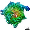
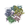
Database: EMDB / ID: EMD-22250
Title
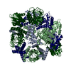
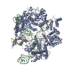
The Heterotetramers of Holin-Antiholin
Map data
sRI-sT heterotetramers
Sample
Complex: The heterotetramers of holin-antiholin
Function / homology
Function and homology information
host cell periplasmic space / pore-forming activity / molecular function inhibitor activity / viral release from host cell by cytolysis / killing of cells of another organism / host cell plasma membrane / DNA binding / membrane Similarity search - Function
Bacteriophage T4, GpT, holin / Bacteriophage T holin / Antiholin T4 type / T4-like virus antiholin family Similarity search - Domain/homology
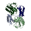
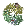
Journal: J Mol Biol / Year: 2020 Title: The Structural Basis of T4 Phage Lysis Control: DNA as the Signal for Lysis Inhibition. Authors: Inna V Krieger / Vladimir Kuznetsov / Jeng-Yih Chang / Junjie Zhang / Samir H Moussa / Ryland F Young / James C Sacchettini / Abstract: Optimal phage propagation depends on the regulation of the lysis of the infected host cell. In T4 phage infection, lysis occurs when the holin protein (T) forms lesions in the host membrane. However, ...Optimal phage propagation depends on the regulation of the lysis of the infected host cell. In T4 phage infection, lysis occurs when the holin protein (T) forms lesions in the host membrane. However, the lethal function of T can be blocked by an antiholin (RI) during lysis inhibition (LIN). LIN sets if the infected cell undergoes superinfection, then the lysis is delayed until host/phage ratio becomes more favorable for the release of progeny. It has been thought that a signal derived from the superinfection is required to activate RI. Here we report structures that suggest a radically different model in which RI binds to T irrespective of superinfection, causing it to accumulate in a membrane as heterotetrameric 2RI-2T complex. Moreover, we show the complex binds non-specifically to DNA, suggesting that the gDNA from the superinfecting phage serves as the LIN signal and that stabilization of the complex by DNA binding is what defines LIN. Finally, we show that soluble domain of free RI crystallizes in a domain-swapped homotetramer, which likely works as a sink for RI molecules released from the RI-T complex to ensure efficient lysis. These results constitute the first structural basis and a new model not only for the historic LIN phenomenon but also for the temporal regulation of phage lysis in general.
History
Deposition
Jun 29, 2020
-
Header (metadata) release
Jul 8, 2020
-
Map release
Jul 8, 2020
-
Update
Aug 12, 2020
-
Current status
Aug 12, 2020
Processing site: RCSB / Status: Released
-
Structure visualization
Movie
Surface view with section colored by density value
In the structure databanks used in Yorodumi, some data are registered as the other names, "COVID-19 virus" and "2019-nCoV". Here are the details of the virus and the list of structure data.
Jan 31, 2019. EMDB accession codes are about to change! (news from PDBe EMDB page)
EMDB accession codes are about to change! (news from PDBe EMDB page)
The allocation of 4 digits for EMDB accession codes will soon come to an end. Whilst these codes will remain in use, new EMDB accession codes will include an additional digit and will expand incrementally as the available range of codes is exhausted. The current 4-digit format prefixed with “EMD-” (i.e. EMD-XXXX) will advance to a 5-digit format (i.e. EMD-XXXXX), and so on. It is currently estimated that the 4-digit codes will be depleted around Spring 2019, at which point the 5-digit format will come into force.
The EM Navigator/Yorodumi systems omit the EMD- prefix.
Related info.:Q: What is EMD? / ID/Accession-code notation in Yorodumi/EM Navigator
Yorodumi is a browser for structure data from EMDB, PDB, SASBDB, etc.
This page is also the successor to EM Navigator detail page, and also detail information page/front-end page for Omokage search.
The word "yorodu" (or yorozu) is an old Japanese word meaning "ten thousand". "mi" (miru) is to see.
Related info.:EMDB / PDB / SASBDB / Comparison of 3 databanks / Yorodumi Search / Aug 31, 2016. New EM Navigator & Yorodumi / Yorodumi Papers / Jmol/JSmol / Function and homology information / Changes in new EM Navigator and Yorodumi
 Movie
Movie Controller


 Open data
 Basic information
 Map data
 Sample
 Function and homology information
 Escherichia phage T4 (virus)
 Authors
 United States, 1 items
  Citation
 Structure visualization
 Downloads & links
 emd_22250.png
 http://ftp.pdbj.org/pub/emdb/structures/EMD-22250


 Z (Sec.)
 Y (Row.)
 X (Col.)


 Sample components
 Processing
 Electron microscopy
 FIELD EMISSION GUN