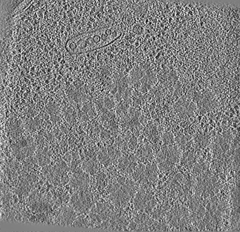
- EMDB-21953: Downsampled and filtered tomogram of a region from a cryoFIB-SEM-... -
+
データを開く
IDまたはキーワード:
読み込み中...
-
基本情報
登録情報
データベース: EMDB / ID: EMD-21953
タイトル
Downsampled and filtered tomogram of a region from a cryoFIB-SEM-generated lamellae of yeast cells under heat shock stress showing large protein aggregates
マップデータ
Binned-by-8 and filtered tomogram of a region from a cryoFIB-SEM-generated lamellae of yeast cells under heat shock stress showing large protein aggregates.
試料
細胞: Downsamples and filtered tomogram of a region from a cryoFIB-SEM-generated lamellae of yeast cells under heat shock stress showing large protein aggregates
National Institutes of Health/National Institute of Neurological Disorders and Stroke (NIH/NINDS)
P01NS092525
米国
National Institutes of Health/National Institute of General Medical Sciences (NIH/NIGMS)
5P41GM103832
米国
National Institutes of Health/National Institute on Aging (NIH/NIA)
PO1AG054407
米国
National Institutes of Health/National Institute of Neurological Disorders and Stroke (NIH/NINDS)
F32NS086253
米国
National Institutes of Health/National Institute of General Medical Sciences (NIH/NIGMS)
S10OD021600
米国
Department of Energy (DOE, United States)
BERFWP 100463
米国
引用
ジャーナル: Structure / 年: 2020 タイトル: Multi-scale 3D Cryo-Correlative Microscopy for Vitrified Cells. 著者: Gong-Her Wu / Patrick G Mitchell / Jesus G Galaz-Montoya / Corey W Hecksel / Emily M Sontag / Vimal Gangadharan / Jeffrey Marshman / David Mankus / Margaret E Bisher / Abigail K R Lytton-Jean ...著者: Gong-Her Wu / Patrick G Mitchell / Jesus G Galaz-Montoya / Corey W Hecksel / Emily M Sontag / Vimal Gangadharan / Jeffrey Marshman / David Mankus / Margaret E Bisher / Abigail K R Lytton-Jean / Judith Frydman / Kirk Czymmek / Wah Chiu / 要旨: Three-dimensional (3D) visualization of vitrified cells can uncover structures of subcellular complexes without chemical fixation or staining. Here, we present a pipeline integrating three imaging ...Three-dimensional (3D) visualization of vitrified cells can uncover structures of subcellular complexes without chemical fixation or staining. Here, we present a pipeline integrating three imaging modalities to visualize the same specimen at cryogenic temperature at different scales: cryo-fluorescence confocal microscopy, volume cryo-focused ion beam scanning electron microscopy, and transmission cryo-electron tomography. Our proof-of-concept benchmark revealed the 3D distribution of organelles and subcellular structures in whole heat-shocked yeast cells, including the ultrastructure of protein inclusions that recruit fluorescently-labeled chaperone Hsp104. Since our workflow efficiently integrates imaging at three different scales and can be applied to other types of cells, it could be used for large-scale phenotypic studies of frozen-hydrated specimens in a variety of healthy and diseased conditions with and without treatments.
ダウンロード / ファイル: emd_21953.map.gz / 形式: CCP4 / 大きさ: 174.3 MB / タイプ: IMAGE STORED AS FLOATING POINT NUMBER (4 BYTES)
注釈
Binned-by-8 and filtered tomogram of a region from a cryoFIB-SEM-generated lamellae of yeast cells under heat shock stress showing large protein aggregates.
A: 13251.054 Å / B: 12808.43 Å / C: 5698.783 Å α=β=γ: 90.0 °
CCP4マップ ヘッダ情報:
mode
Image stored as Reals
Å/pix. X/Y/Z
27.663995824635
27.663995680346
27.663995145631
M x/y/z
479
463
206
origin x/y/z
0.000
0.000
0.000
length x/y/z
13251.054
12808.430
5698.783
α/β/γ
90.000
90.000
90.000
start NX/NY/NZ
79
74
0
NX/NY/NZ
93
103
213
MAP C/R/S
1
2
3
start NC/NR/NS
0
0
-103
NC/NR/NS
479
463
206
D min/max/mean
-3.000
3.000
-0.000
-
添付データ
-
試料の構成要素
-
全体 : Downsamples and filtered tomogram of a region from a cryoFIB-SEM-...
全体
名称: Downsamples and filtered tomogram of a region from a cryoFIB-SEM-generated lamellae of yeast cells under heat shock stress showing large protein aggregates
要素
細胞: Downsamples and filtered tomogram of a region from a cryoFIB-SEM-generated lamellae of yeast cells under heat shock stress showing large protein aggregates
-
超分子 #1: Downsamples and filtered tomogram of a region from a cryoFIB-SEM-...
超分子
名称: Downsamples and filtered tomogram of a region from a cryoFIB-SEM-generated lamellae of yeast cells under heat shock stress showing large protein aggregates タイプ: cell / ID: 1 / 親要素: 0
由来(天然)
生物種: Saccharomyces cerevisiae (パン酵母) / 株: BY4741
-
実験情報
-
構造解析
手法
クライオ電子顕微鏡法
解析
電子線トモグラフィー法
試料の集合状態
cell
-
試料調製
緩衝液
pH: 7
凍結
凍結剤: ETHANE
切片作成
集束イオンビーム - 装置: OTHER / 集束イオンビーム - イオン: OTHER / 集束イオンビーム - 電圧: 30 kV / 集束イオンビーム - 電流: 20 nA / 集束イオンビーム - 時間: 60 sec. / 集束イオンビーム - 温度: 100 K / 集束イオンビーム - Initial thickness: 1000 nm / 集束イオンビーム - 最終 厚さ: 520 nm 集束イオンビーム - 詳細: The value given for _emd_sectioning_focused_ion_beam.instrument is Zeiss Crossbeam 540 FIB-SEM. This is not in a list of allowed values set(['DB235', 'OTHER']) so ...集束イオンビーム - 詳細: The value given for _emd_sectioning_focused_ion_beam.instrument is Zeiss Crossbeam 540 FIB-SEM. This is not in a list of allowed values set(['DB235', 'OTHER']) so OTHER is written into the XML file.
アルゴリズム: SIMULTANEOUS ITERATIVE (SIRT) / ソフトウェア - 名称: IMOD 詳細: The SIRT, downsampled, and filtered tomogram was computed for visualization only. The related EMDB entry showcasing a ribosome subtomogram average used similar tomograms but reconstructed ...詳細: The SIRT, downsampled, and filtered tomogram was computed for visualization only. The related EMDB entry showcasing a ribosome subtomogram average used similar tomograms but reconstructed with weighted-back-projection, at full-size, and unfiltered. 使用した粒子像数: 121
 ムービー
ムービー コントローラー


 データを開く
 基本情報
基本情報 マップデータ
 試料
試料
 データ登録者
 米国, 6件
  引用
引用 構造の表示
構造の表示 ムービービューア
 ダウンロードとリンク
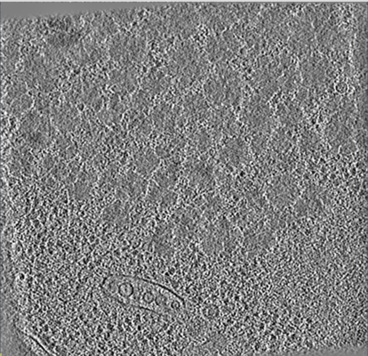 emd_21953.png
 http://ftp.pdbj.org/pub/emdb/structures/EMD-21953
 Z (Sec.)
 Y (Row.)
 X (Col.)


 試料の構成要素
 解析
解析 電子顕微鏡法
 FIELD EMISSION GUN
