+ データを開く
- 基本情報
基本情報
| 登録情報 | データベース: EMDB / ID: EMD-21925 | |||||||||
|---|---|---|---|---|---|---|---|---|---|---|
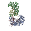
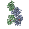
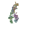
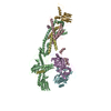
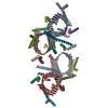
| タイトル | Structural basis of alphaE-catenin - F-actin catch bond behavior | |||||||||
 マップデータ マップデータ | alphaE-catenin - F-actin catch bond behavior | |||||||||
 試料 試料 |
| |||||||||
 キーワード キーワード | alphaE-catenin / catch bond / adherens junction / CELL ADHESION | |||||||||
| 機能・相同性 |  機能・相同性情報 機能・相同性情報negative regulation of integrin-mediated signaling pathway / VEGFR2 mediated vascular permeability / Adherens junctions interactions / RHO GTPases activate IQGAPs / gamma-catenin binding / epithelial cell-cell adhesion / zonula adherens / gap junction assembly / cellular response to indole-3-methanol / flotillin complex ...negative regulation of integrin-mediated signaling pathway / VEGFR2 mediated vascular permeability / Adherens junctions interactions / RHO GTPases activate IQGAPs / gamma-catenin binding / epithelial cell-cell adhesion / zonula adherens / gap junction assembly / cellular response to indole-3-methanol / flotillin complex / vinculin binding / Myogenesis / catenin complex / apical junction assembly / negative regulation of cell motility / positive regulation of extrinsic apoptotic signaling pathway in absence of ligand / positive regulation of smoothened signaling pathway / negative regulation of protein localization to nucleus / axon regeneration / cytoskeletal motor activator activity / negative regulation of neuroblast proliferation / smoothened signaling pathway / myosin heavy chain binding / tropomyosin binding / actin filament bundle / establishment or maintenance of cell polarity / troponin I binding / filamentous actin / odontogenesis of dentin-containing tooth / mesenchyme migration / actin filament bundle assembly / skeletal muscle myofibril / striated muscle thin filament / skeletal muscle thin filament assembly / intercalated disc / actin monomer binding / neuroblast proliferation / negative regulation of extrinsic apoptotic signaling pathway in absence of ligand / ovarian follicle development / skeletal muscle fiber development / stress fiber / titin binding / actin filament polymerization / extrinsic apoptotic signaling pathway in absence of ligand / acrosomal vesicle / integrin-mediated signaling pathway / adherens junction / actin filament / filopodium / cell motility / beta-catenin binding / 加水分解酵素; 酸無水物に作用; 酸無水物に作用・細胞または細胞小器官の運動に関与 / response to estrogen / male gonad development / calcium-dependent protein binding / actin filament binding / intracellular protein localization / actin cytoskeleton / lamellipodium / regulation of cell population proliferation / cell body / hydrolase activity / cadherin binding / protein domain specific binding / apoptotic process / calcium ion binding / positive regulation of gene expression / negative regulation of apoptotic process / structural molecule activity / magnesium ion binding / Golgi apparatus / ATP binding / identical protein binding / nucleus / plasma membrane / cytoplasm 類似検索 - 分子機能 | |||||||||
| 生物種 |   | |||||||||
| 手法 | らせん対称体再構成法 / クライオ電子顕微鏡法 / 解像度: 3.56 Å | |||||||||
 データ登録者 データ登録者 | Xu XP / Pokutta S | |||||||||
| 資金援助 |  米国, 1件 米国, 1件
| |||||||||
 引用 引用 |  ジャーナル: Elife / 年: 2020 ジャーナル: Elife / 年: 2020タイトル: Structural basis of αE-catenin-F-actin catch bond behavior. 著者: Xiao-Ping Xu / Sabine Pokutta / Megan Torres / Mark F Swift / Dorit Hanein / Niels Volkmann / William I Weis /   要旨: Cell-cell and cell-matrix junctions transmit mechanical forces during tissue morphogenesis and homeostasis. α-Catenin links cell-cell adhesion complexes to the actin cytoskeleton, and mechanical ...Cell-cell and cell-matrix junctions transmit mechanical forces during tissue morphogenesis and homeostasis. α-Catenin links cell-cell adhesion complexes to the actin cytoskeleton, and mechanical load strengthens its binding to F-actin in a direction-sensitive manner. Specifically, optical trap experiments revealed that force promotes a transition between weak and strong actin-bound states. Here, we describe the cryo-electron microscopy structure of the F-actin-bound αE-catenin actin-binding domain, which in solution forms a five-helix bundle. In the actin-bound structure, the first helix of the bundle dissociates and the remaining four helices and connecting loops rearrange to form the interface with actin. Deletion of the first helix produces strong actin binding in the absence of force, suggesting that the actin-bound structure corresponds to the strong state. Our analysis explains how mechanical force applied to αE-catenin or its homolog vinculin favors the strongly bound state, and the dependence of catch bond strength on the direction of applied force. | |||||||||
| 履歴 |
|
- 構造の表示
構造の表示
| ムービー |
 ムービービューア ムービービューア |
|---|---|
| 構造ビューア | EMマップ:  SurfView SurfView Molmil Molmil Jmol/JSmol Jmol/JSmol |
| 添付画像 |
- ダウンロードとリンク
-EMDBアーカイブ
| マップデータ |  emd_21925.map.gz emd_21925.map.gz | 6.1 MB |  EMDBマップデータ形式 EMDBマップデータ形式 | |
|---|---|---|---|---|
| ヘッダ (付随情報) |  emd-21925-v30.xml emd-21925-v30.xml emd-21925.xml emd-21925.xml | 18.2 KB 18.2 KB | 表示 表示 |  EMDBヘッダ EMDBヘッダ |
| 画像 |  emd_21925.png emd_21925.png | 276.4 KB | ||
| Filedesc metadata |  emd-21925.cif.gz emd-21925.cif.gz | 6.8 KB | ||
| アーカイブディレクトリ |  http://ftp.pdbj.org/pub/emdb/structures/EMD-21925 http://ftp.pdbj.org/pub/emdb/structures/EMD-21925 ftp://ftp.pdbj.org/pub/emdb/structures/EMD-21925 ftp://ftp.pdbj.org/pub/emdb/structures/EMD-21925 | HTTPS FTP |
-検証レポート
| 文書・要旨 |  emd_21925_validation.pdf.gz emd_21925_validation.pdf.gz | 480.3 KB | 表示 |  EMDB検証レポート EMDB検証レポート |
|---|---|---|---|---|
| 文書・詳細版 |  emd_21925_full_validation.pdf.gz emd_21925_full_validation.pdf.gz | 479.9 KB | 表示 | |
| XML形式データ |  emd_21925_validation.xml.gz emd_21925_validation.xml.gz | 5.9 KB | 表示 | |
| CIF形式データ |  emd_21925_validation.cif.gz emd_21925_validation.cif.gz | 6.7 KB | 表示 | |
| アーカイブディレクトリ |  https://ftp.pdbj.org/pub/emdb/validation_reports/EMD-21925 https://ftp.pdbj.org/pub/emdb/validation_reports/EMD-21925 ftp://ftp.pdbj.org/pub/emdb/validation_reports/EMD-21925 ftp://ftp.pdbj.org/pub/emdb/validation_reports/EMD-21925 | HTTPS FTP |
-関連構造データ
- リンク
リンク
| EMDBのページ |  EMDB (EBI/PDBe) / EMDB (EBI/PDBe) /  EMDataResource EMDataResource |
|---|---|
| 「今月の分子」の関連する項目 |
- マップ
マップ
| ファイル |  ダウンロード / ファイル: emd_21925.map.gz / 形式: CCP4 / 大きさ: 17.1 MB / タイプ: IMAGE STORED AS FLOATING POINT NUMBER (4 BYTES) ダウンロード / ファイル: emd_21925.map.gz / 形式: CCP4 / 大きさ: 17.1 MB / タイプ: IMAGE STORED AS FLOATING POINT NUMBER (4 BYTES) | ||||||||||||||||||||||||||||||||||||||||||||||||||||||||||||||||||||
|---|---|---|---|---|---|---|---|---|---|---|---|---|---|---|---|---|---|---|---|---|---|---|---|---|---|---|---|---|---|---|---|---|---|---|---|---|---|---|---|---|---|---|---|---|---|---|---|---|---|---|---|---|---|---|---|---|---|---|---|---|---|---|---|---|---|---|---|---|---|
| 注釈 | alphaE-catenin - F-actin catch bond behavior | ||||||||||||||||||||||||||||||||||||||||||||||||||||||||||||||||||||
| 投影像・断面図 | 画像のコントロール
画像は Spider により作成 | ||||||||||||||||||||||||||||||||||||||||||||||||||||||||||||||||||||
| ボクセルのサイズ | X=Y=Z: 1.035 Å | ||||||||||||||||||||||||||||||||||||||||||||||||||||||||||||||||||||
| 密度 |
| ||||||||||||||||||||||||||||||||||||||||||||||||||||||||||||||||||||
| 対称性 | 空間群: 1 | ||||||||||||||||||||||||||||||||||||||||||||||||||||||||||||||||||||
| 詳細 | EMDB XML:
CCP4マップ ヘッダ情報:
| ||||||||||||||||||||||||||||||||||||||||||||||||||||||||||||||||||||
-添付データ
- 試料の構成要素
-全体 : Complex of alphaE-catenin actin binding domain with F-actin
| 全体 | 名称: Complex of alphaE-catenin actin binding domain with F-actin |
|---|---|
| 要素 |
|
-超分子 #1: Complex of alphaE-catenin actin binding domain with F-actin
| 超分子 | 名称: Complex of alphaE-catenin actin binding domain with F-actin タイプ: complex / ID: 1 / 親要素: 0 / 含まれる分子: #1-#2 |
|---|
-超分子 #2: Actin, alpha skeletal muscle
| 超分子 | 名称: Actin, alpha skeletal muscle / タイプ: complex / ID: 2 / 親要素: 1 / 含まれる分子: #1 |
|---|---|
| 由来(天然) | 生物種:  |
-超分子 #3: Catenin alpha-1
| 超分子 | 名称: Catenin alpha-1 / タイプ: complex / ID: 3 / 親要素: 1 / 含まれる分子: #2 |
|---|---|
| 由来(天然) | 生物種:  |
-分子 #1: Actin, alpha skeletal muscle
| 分子 | 名称: Actin, alpha skeletal muscle / タイプ: protein_or_peptide / ID: 1 / コピー数: 6 / 光学異性体: LEVO |
|---|---|
| 由来(天然) | 生物種:  |
| 分子量 | 理論値: 42.109973 KDa |
| 配列 | 文字列: MCDEDETTAL VCDNGSGLVK AGFAGDDAPR AVFPSIVGRP RHQGVMVGMG QKDSYVGDEA QSKRGILTLK YPIE(HIC)G IIT NWDDMEKIWH HTFYNELRVA PEEHPTLLTE APLNPKANRE KMTQIMFETF NVPAMYVAIQ AVLSLYASGR TTGIVLD SG ...文字列: MCDEDETTAL VCDNGSGLVK AGFAGDDAPR AVFPSIVGRP RHQGVMVGMG QKDSYVGDEA QSKRGILTLK YPIE(HIC)G IIT NWDDMEKIWH HTFYNELRVA PEEHPTLLTE APLNPKANRE KMTQIMFETF NVPAMYVAIQ AVLSLYASGR TTGIVLD SG DGVTHNVPIY EGYALPHAIM RLDLAGRDLT DYLMKILTER GYSFVTTAER EIVRDIKEKL CYVALDFENE MATAASSS S LEKSYELPDG QVITIGNERF RCPETLFQPS FIGMESAGIH ETTYNSIMKC DIDIRKDLYA NNVMSGGTTM YPGIADRMQ KEITALAPST MKIKIIAPPE RKYSVWIGGS ILASLSTFQQ MWITKQEYDE AGPSIVHRKC F UniProtKB: Actin, alpha skeletal muscle |
-分子 #2: Catenin alpha-1
| 分子 | 名称: Catenin alpha-1 / タイプ: protein_or_peptide / ID: 2 / コピー数: 6 / 光学異性体: LEVO |
|---|---|
| 由来(天然) | 生物種:  |
| 分子量 | 理論値: 25.999281 KDa |
| 組換発現 | 生物種:  |
| 配列 | 文字列: AIMAQLPQEQ KAKIAEQVAS FQEEKSKLDA EVSKWDDSGN DIIVLAKQMC MIMMEMTDFT RGKGPLKNTS DVISAAKKIA EAGSRMDKL GRTIADHCPD SACKQDLLAY LQRIALYCHQ LNICSKVKAE VQNLGGELVV SGVDSAMSLI QAAKNLMNAV V QTVKASYV ...文字列: AIMAQLPQEQ KAKIAEQVAS FQEEKSKLDA EVSKWDDSGN DIIVLAKQMC MIMMEMTDFT RGKGPLKNTS DVISAAKKIA EAGSRMDKL GRTIADHCPD SACKQDLLAY LQRIALYCHQ LNICSKVKAE VQNLGGELVV SGVDSAMSLI QAAKNLMNAV V QTVKASYV ASTKYQKSQG MASLNLPAVS WKMKAPEKKP LVKREKQDET QTKIKRASQK KHVNPVQALS EFKAMDSI UniProtKB: Catenin alpha-1 |
-分子 #3: MAGNESIUM ION
| 分子 | 名称: MAGNESIUM ION / タイプ: ligand / ID: 3 / コピー数: 6 / 式: MG |
|---|---|
| 分子量 | 理論値: 24.305 Da |
-分子 #4: ADENOSINE-5'-DIPHOSPHATE
| 分子 | 名称: ADENOSINE-5'-DIPHOSPHATE / タイプ: ligand / ID: 4 / コピー数: 6 / 式: ADP |
|---|---|
| 分子量 | 理論値: 427.201 Da |
| Chemical component information |  ChemComp-ADP: |
-実験情報
-構造解析
| 手法 | クライオ電子顕微鏡法 |
|---|---|
 解析 解析 | らせん対称体再構成法 |
| 試料の集合状態 | helical array |
- 試料調製
試料調製
| 緩衝液 | pH: 7.5 |
|---|---|
| 凍結 | 凍結剤: ETHANE |
- 電子顕微鏡法
| 顕微鏡 | FEI TITAN KRIOS |
|---|---|
| 撮影 | フィルム・検出器のモデル: FEI FALCON II (4k x 4k) 検出モード: INTEGRATING / デジタル化 - サイズ - 横: 4096 pixel / デジタル化 - サイズ - 縦: 4096 pixel / デジタル化 - 画像ごとのフレーム数: 2-7 / 撮影したグリッド数: 3 / 実像数: 5573 / 平均露光時間: 1.0 sec. / 平均電子線量: 60.0 e/Å2 |
| 電子線 | 加速電圧: 300 kV / 電子線源:  FIELD EMISSION GUN FIELD EMISSION GUN |
| 電子光学系 | 照射モード: FLOOD BEAM / 撮影モード: BRIGHT FIELD / Cs: 2.7 mm 最大 デフォーカス(公称値): 2.8000000000000003 µm 最小 デフォーカス(公称値): 0.8 µm / 倍率(公称値): 75000 |
| 試料ステージ | 試料ホルダーモデル: FEI TITAN KRIOS AUTOGRID HOLDER ホルダー冷却材: NITROGEN |
| 実験機器 |  モデル: Titan Krios / 画像提供: FEI Company |
- 画像解析
画像解析
| 最終 再構成 | 想定した対称性 - らせんパラメータ - Δz: 27.4 Å 想定した対称性 - らせんパラメータ - ΔΦ: -166.9 ° 想定した対称性 - らせんパラメータ - 軸対称性: C1 (非対称) 解像度のタイプ: BY AUTHOR / 解像度: 3.56 Å / 解像度の算出法: FSC 0.143 CUT-OFF / ソフトウェア - 名称: RELION (ver. 3) / 使用した粒子像数: 422822 |
|---|---|
| Segment selection | 選択した数: 728331 |
| 初期モデル | モデルのタイプ: OTHER 詳細: in-house rabbit skeletal actin filament reconstruction filtered to 4 nm |
| 最終 角度割当 | タイプ: NOT APPLICABLE / ソフトウェア - 名称: RELION (ver. 3) |
 ムービー
ムービー コントローラー




 Z (Sec.)
 Y (Row.)
 X (Col.)


