 Movie
Movie Controller


+ Open data

- Basic information
| Entry | Database: EMDB / ID: EMD-2169 | |||||||||
|---|---|---|---|---|---|---|---|---|---|---|
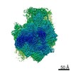
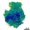
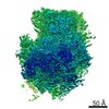
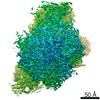
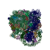
| Title | Cryo-EM structure of the 60S-Arx1-Rei1 complex | |||||||||
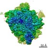
 Map data Map data | Cryo-EM reconstruction of the 60S-Arx1-Rei1 complex | |||||||||
 Sample Sample |
| |||||||||
 Keywords Keywords | large ribosomal subunit / ribosome biogenesis / ribosome maturation factor | |||||||||
| Function / homology |  Function and homology information Function and homology informationHydrolases / Ribosome Quality Control (RQC) complex extracts and degrades nascent peptide / PELO:HBS1L and ABCE1 dissociate a ribosome on a non-stop mRNA / pre-mRNA 5'-splice site binding / cytosolic large ribosomal subunit assembly / response to cycloheximide / cleavage in ITS2 between 5.8S rRNA and LSU-rRNA of tricistronic rRNA transcript (SSU-rRNA, 5.8S rRNA, LSU-rRNA) / SRP-dependent cotranslational protein targeting to membrane / GTP hydrolysis and joining of the 60S ribosomal subunit / Formation of a pool of free 40S subunits ...Hydrolases / Ribosome Quality Control (RQC) complex extracts and degrades nascent peptide / PELO:HBS1L and ABCE1 dissociate a ribosome on a non-stop mRNA / pre-mRNA 5'-splice site binding / cytosolic large ribosomal subunit assembly / response to cycloheximide / cleavage in ITS2 between 5.8S rRNA and LSU-rRNA of tricistronic rRNA transcript (SSU-rRNA, 5.8S rRNA, LSU-rRNA) / SRP-dependent cotranslational protein targeting to membrane / GTP hydrolysis and joining of the 60S ribosomal subunit / Formation of a pool of free 40S subunits / preribosome, large subunit precursor / Nonsense Mediated Decay (NMD) independent of the Exon Junction Complex (EJC) / Nonsense Mediated Decay (NMD) enhanced by the Exon Junction Complex (EJC) / negative regulation of mRNA splicing, via spliceosome / L13a-mediated translational silencing of Ceruloplasmin expression / translational elongation / ribosomal large subunit export from nucleus / protein kinase activator activity / ribonucleoprotein complex binding / 90S preribosome / translational termination / regulation of translational fidelity / maturation of LSU-rRNA / protein-RNA complex assembly / Neutrophil degranulation / ribosomal large subunit biogenesis / maturation of LSU-rRNA from tricistronic rRNA transcript (SSU-rRNA, 5.8S rRNA, LSU-rRNA) / macroautophagy / translational initiation / maintenance of translational fidelity / modification-dependent protein catabolic process / metallopeptidase activity / protein tag activity / rRNA processing / ribosome biogenesis / 5S rRNA binding / ribosomal large subunit assembly / large ribosomal subunit rRNA binding / cytosolic large ribosomal subunit / cytoplasmic translation / negative regulation of translation / rRNA binding / structural constituent of ribosome / protein ubiquitination / ribosome / translation / response to antibiotic / mRNA binding / ubiquitin protein ligase binding / nucleolus / proteolysis / RNA binding / zinc ion binding / nucleoplasm / metal ion binding / nucleus / cytosol / cytoplasm Similarity search - Function | |||||||||
| Biological species |  | |||||||||
| Method | single particle reconstruction / cryo EM / negative staining / Resolution: 8.1 Å | |||||||||
 Authors Authors | Greber BJ / Boehringer D / Montellese C / Ban N | |||||||||
 Citation Citation | Journal: Nat Struct Mol Biol / Year: 2012 Title: Cryo-EM structures of Arx1 and maturation factors Rei1 and Jjj1 bound to the 60S ribosomal subunit. Authors: Basil J Greber / Daniel Boehringer / Christian Montellese / Nenad Ban /  Abstract: Eukaryotic ribosome biogenesis requires many protein factors that facilitate the assembly, nuclear export and final maturation of 40S and 60S particles. We have biochemically characterized ribosomal ...Eukaryotic ribosome biogenesis requires many protein factors that facilitate the assembly, nuclear export and final maturation of 40S and 60S particles. We have biochemically characterized ribosomal complexes of the yeast 60S-biogenesis factor Arx1 and late-maturation factors Rei1 and Jjj1 and determined their cryo-EM structures. Arx1 was visualized bound to the 60S subunit together with Rei1, at 8.1-Å resolution, to reveal the molecular details of Arx1 binding whereby Arx1 arrests the eukaryotic-specific rRNA expansion segment 27 near the polypeptide tunnel exit. Rei1 and Jjj1, which have been implicated in Arx1 recycling, bind in the vicinity of Arx1 and form a network of interactions. We suggest that, in addition to the role of Arx1 during pre-60S nuclear export, the binding of Arx1 conformationally locks the pre-60S subunit and inhibits the premature association of nascent chain-processing factors to the polypeptide tunnel exit. | |||||||||
| History |
|
- Structure visualization
| Movie |
Movie viewer |
|---|---|
| Structure viewer | EM map: SurfViewMolmilJmol/JSmol |
| Supplemental images |

- Downloads & links
-EMDB archive
| Map data | emd_2169.map.gz | 37.6 MB | EMDB map data format | |
|---|---|---|---|---|
| Header (meta data) | emd-2169-v30.xmlemd-2169.xml | 16.8 KB 16.8 KB | Display Display | EMDB header |
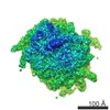
| Images | 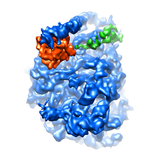 EMD-2169.jpg EMD-2169.jpg | 129 KB | ||
| Archive directory |  http://ftp.pdbj.org/pub/emdb/structures/EMD-2169ftp://ftp.pdbj.org/pub/emdb/structures/EMD-2169 http://ftp.pdbj.org/pub/emdb/structures/EMD-2169ftp://ftp.pdbj.org/pub/emdb/structures/EMD-2169 | HTTPS FTP |
-Related structure data
| Related structure data |  4v8tMC 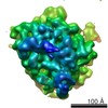 2167C 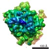 2168C M: atomic model generated by this map C: citing same article ( |
|---|---|
| Similar structure data |

-Links
| EMDB pages | EMDB (EBI/PDBe) / EMDataResource |
|---|---|
| Related items in Molecule of the Month |


-Map
| File | Download / File: emd_2169.map.gz / Format: CCP4 / Size: 41.9 MB / Type: IMAGE STORED AS FLOATING POINT NUMBER (4 BYTES) | ||||||||||||||||||||||||||||||||||||||||||||||||||||||||||||||||||||
|---|---|---|---|---|---|---|---|---|---|---|---|---|---|---|---|---|---|---|---|---|---|---|---|---|---|---|---|---|---|---|---|---|---|---|---|---|---|---|---|---|---|---|---|---|---|---|---|---|---|---|---|---|---|---|---|---|---|---|---|---|---|---|---|---|---|---|---|---|---|
| Annotation | Cryo-EM reconstruction of the 60S-Arx1-Rei1 complex | ||||||||||||||||||||||||||||||||||||||||||||||||||||||||||||||||||||
| Projections & slices | Image control
Images are generated by Spider. | ||||||||||||||||||||||||||||||||||||||||||||||||||||||||||||||||||||
| Voxel size | X=Y=Z: 1.81 Å | ||||||||||||||||||||||||||||||||||||||||||||||||||||||||||||||||||||
| Density |
| ||||||||||||||||||||||||||||||||||||||||||||||||||||||||||||||||||||
| Symmetry | Space group: 1 | ||||||||||||||||||||||||||||||||||||||||||||||||||||||||||||||||||||
| Details | EMDB XML:
CCP4 map header:
| ||||||||||||||||||||||||||||||||||||||||||||||||||||||||||||||||||||
 Z (Sec.)
 Y (Row.)
 X (Col.)


-Supplemental data
- Sample components
-Entire : 60S ribosomal subunit in complex with Arx1 and Rei1
| Entire | Name: 60S ribosomal subunit in complex with Arx1 and Rei1 |
|---|---|
| Components |
|
-Supramolecule #1000: 60S ribosomal subunit in complex with Arx1 and Rei1
| Supramolecule | Name: 60S ribosomal subunit in complex with Arx1 and Rei1 / type: sample / ID: 1000 / Oligomeric state: One monomer each of 60S, Arx1, and Rei1 / Number unique components: 3 |
|---|---|
| Molecular weight | Theoretical: 2.3 MDa |
-Supramolecule #1: 60S ribosomal subunit
| Supramolecule | Name: 60S ribosomal subunit / type: complex / ID: 1 / Name.synonym: 60S / Recombinant expression: No / Ribosome-details: ribosome-eukaryote: LSU 60S |
|---|---|
| Source (natural) | Organism: |
| Molecular weight | Theoretical: 2.2 MDa |
-Macromolecule #1: Arx1
| Macromolecule | Name: Arx1 / type: protein_or_peptide / ID: 1 / Details: N-terminal His-tag / Number of copies: 1 / Oligomeric state: monomer / Recombinant expression: Yes |
|---|---|
| Source (natural) | Organism: |
| Molecular weight | Theoretical: 65 KDa |
| Recombinant expression | Organism:  |
| Sequence | InterPro: Peptidase M24 |
-Macromolecule #2: Rei1
| Macromolecule | Name: Rei1 / type: protein_or_peptide / ID: 2 / Details: C-terminal His-tag / Number of copies: 1 / Oligomeric state: monomer / Recombinant expression: Yes |
|---|---|
| Source (natural) | Organism: |
| Molecular weight | Theoretical: 46 KDa |
| Recombinant expression | Organism: |
| Sequence | InterPro: INTERPRO: IPR007087, INTERPRO: IPR015880 |
-Experimental details
-Structure determination
| Method | negative staining, cryo EM |
|---|---|
 Processing Processing | single particle reconstruction |
| Aggregation state | particle |
-Sample preparation
| Buffer | pH: 8 Details: 20 mM Hepes-NaOH pH 8.0, 50 mM NaCl, 5 mM MgCl2, 5 mM beta-mercaptoethanol |
|---|---|
| Staining | Type: NEGATIVE / Details: cryo |
| Grid | Details: Quantifoil holey carbon grid R2/1 |
| Vitrification | Cryogen name: ETHANE / Chamber temperature: 80 K / Instrument: HOMEMADE PLUNGER / Details: Vitrification instrument: manual plunger / Method: manual blotting |
- Electron microscopy #1
| Microscopy ID | 1 |
|---|---|
| Microscope | FEI TECNAI 20 |
| Temperature | Average: 87 K |
| Date | Feb 21, 2012 |
| Image recording | Category: CCD / Film or detector model: GATAN ULTRASCAN 4000 (4k x 4k) / Digitization - Sampling interval: 15 µm / Average electron dose: 20 e/Å2 Details: images were acquired using a 2 x 2 frame spot scan (per hole) using a serial EM script Bits/pixel: 16 |
| Tilt angle min | 0 |
| Tilt angle max | 0 |
| Electron beam | Acceleration voltage: 200 kV / Electron source:  FIELD EMISSION GUN FIELD EMISSION GUN |
| Electron optics | Illumination mode: FLOOD BEAM / Imaging mode: BRIGHT FIELD / Cs: 2.3 mm / Nominal defocus max: 4.5 µm / Nominal defocus min: 1.5 µm / Nominal magnification: 83000 |
| Sample stage | Specimen holder: eucentric / Specimen holder model: GATAN LIQUID NITROGEN |
-Electron microscopy #2
| Microscopy ID | 2 |
|---|---|
| Microscope | FEI TECNAI 20 |
| Temperature | Average: 87 K |
| Date | Mar 6, 2012 |
| Image recording | Category: CCD / Film or detector model: GATAN ULTRASCAN 4000 (4k x 4k) / Digitization - Sampling interval: 15 µm / Average electron dose: 20 e/Å2 Details: images were acquired using a 2 x 2 frame spot scan (per hole) using a serial EM script Bits/pixel: 16 |
| Tilt angle min | 0 |
| Tilt angle max | 0 |
| Electron beam | Acceleration voltage: 200 kV / Electron source: FIELD EMISSION GUN |
| Electron optics | Illumination mode: FLOOD BEAM / Imaging mode: BRIGHT FIELD / Cs: 2.3 mm / Nominal defocus max: 4.5 µm / Nominal defocus min: 1.5 µm / Nominal magnification: 83000 |
| Sample stage | Specimen holder: eucentric / Specimen holder model: GATAN LIQUID NITROGEN |
-Electron microscopy #3
| Microscopy ID | 3 |
|---|---|
| Microscope | FEI TECNAI 20 |
| Temperature | Average: 87 K |
| Date | Mar 20, 2012 |
| Image recording | Category: CCD / Film or detector model: GATAN ULTRASCAN 4000 (4k x 4k) / Digitization - Sampling interval: 15 µm / Average electron dose: 20 e/Å2 Details: images were acquired using a 2 x 2 frame spot scan (per hole) using a serial EM script Bits/pixel: 16 |
| Tilt angle min | 0 |
| Tilt angle max | 0 |
| Electron beam | Acceleration voltage: 200 kV / Electron source: FIELD EMISSION GUN |
| Electron optics | Illumination mode: FLOOD BEAM / Imaging mode: BRIGHT FIELD / Cs: 2.3 mm / Nominal defocus max: 4.5 µm / Nominal defocus min: 1.5 µm / Nominal magnification: 83000 |
| Sample stage | Specimen holder: eucentric / Specimen holder model: GATAN LIQUID NITROGEN |
-Image processing
| CTF correction | Details: per frame |
|---|---|
| Final reconstruction | Applied symmetry - Point group: C1 (asymmetric) / Algorithm: OTHER / Resolution.type: BY AUTHOR / Resolution: 8.1 Å / Resolution method: FSC 0.5 CUT-OFF / Software - Name: SPIDER Details: Fourier amplitudes of the reconstruction were enhanced using the SAXS curve from ribosomes; subsequently, the map was filtered in SPIDER using a butterworth low-pass filter with a filter midpoint of 0.23. Number images used: 84113 |
-Atomic model buiding 1
| Initial model | PDB ID:  3u5h |
|---|---|
| Software | Name: Chimera |
| Details | Protocol: rigid body |
| Refinement | Space: REAL / Protocol: RIGID BODY FIT / Target criteria: correlation |
| Output model | PDB-4v8t: |
-Atomic model buiding 2
| Initial model | PDB ID: 3u5i |
|---|---|
| Software | Name: Chimera |
| Details | Protocol: rigid body |
| Refinement | Space: REAL / Protocol: RIGID BODY FIT / Target criteria: correlation |
| Output model | PDB-4v8t: |

