 Movie
Movie Controller

[English] 日本語
 Yorodumi
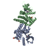
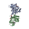
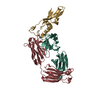
- EMDB-21488: Human diacylglycerol O-acyltransferase 1 complex with oleoyl-CoA ... -
+ Open data

- Basic information
| Entry | Database: EMDB / ID: EMD-21488 | |||||||||||||||
|---|---|---|---|---|---|---|---|---|---|---|---|---|---|---|---|---|
| Title | Human diacylglycerol O-acyltransferase 1 complex with oleoyl-CoA that shows a cleaved acyl-CoA signal | |||||||||||||||
 Map data Map data | Human diacylglycerol O-acyltransferase 1 complexed with oleoyl-CoA showing broken acyl-CoA density | |||||||||||||||
 Sample Sample |
| |||||||||||||||
| Biological species |  Homo sapiens (human) Homo sapiens (human) | |||||||||||||||
| Method | single particle reconstruction / cryo EM / Resolution: 2.8 Å | |||||||||||||||
 Authors Authors | Sui X / Wang K / Gluchowski N / Liao M / Walther CT | |||||||||||||||
| Funding support |  United States, 4 items United States, 4 items
| |||||||||||||||
 Citation Citation | Journal: Nature / Year: 2020 Title: Structure and catalytic mechanism of a human triacylglycerol-synthesis enzyme. Authors: Xuewu Sui / Kun Wang / Nina L Gluchowski / Shane D Elliott / Maofu Liao / Tobias C Walther / Robert V Farese / Abstract: Triacylglycerols store metabolic energy in organisms and have industrial uses as foods and fuels. Excessive accumulation of triacylglycerols in humans causes obesity and is associated with metabolic ...Triacylglycerols store metabolic energy in organisms and have industrial uses as foods and fuels. Excessive accumulation of triacylglycerols in humans causes obesity and is associated with metabolic diseases. Triacylglycerol synthesis is catalysed by acyl-CoA diacylglycerol acyltransferase (DGAT) enzymes, the structures and catalytic mechanisms of which remain unknown. Here we determined the structure of dimeric human DGAT1, a member of the membrane-bound O-acyltransferase (MBOAT) family, by cryo-electron microscopy at approximately 3.0 Å resolution. DGAT1 forms a homodimer through N-terminal segments and a hydrophobic interface, with putative active sites within the membrane region. A structure obtained with oleoyl-CoA substrate resolved at approximately 3.2 Å shows that the CoA moiety binds DGAT1 on the cytosolic side and the acyl group lies deep within a hydrophobic channel, positioning the acyl-CoA thioester bond near an invariant catalytic histidine residue. The reaction centre is located inside a large cavity, which opens laterally to the membrane bilayer, providing lipid access to the active site. A lipid-like density-possibly representing an acyl-acceptor molecule-is located within the reaction centre, orthogonal to acyl-CoA. Insights provided by the DGAT1 structures, together with mutagenesis and functional studies, provide the basis for a model of the catalysis of triacylglycerol synthesis by DGAT. | |||||||||||||||
| History |
|
- Structure visualization
| Movie |
 Movie viewer Movie viewer |
|---|---|
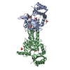
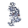
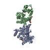
| Structure viewer | EM map: SurfViewMolmilJmol/JSmol |
| Supplemental images |

- Downloads & links
-EMDB archive
| Map data | emd_21488.map.gz | 59.9 MB | EMDB map data format | |
|---|---|---|---|---|
| Header (meta data) | emd-21488-v30.xmlemd-21488.xml | 10.9 KB 10.9 KB | Display Display | EMDB header |
| Images | 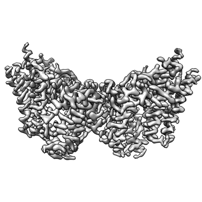 emd_21488.png emd_21488.png | 55.4 KB | ||
| Archive directory |  http://ftp.pdbj.org/pub/emdb/structures/EMD-21488ftp://ftp.pdbj.org/pub/emdb/structures/EMD-21488 http://ftp.pdbj.org/pub/emdb/structures/EMD-21488ftp://ftp.pdbj.org/pub/emdb/structures/EMD-21488 | HTTPS FTP |
-Validation report
| Summary document | emd_21488_validation.pdf.gz | 78 KB | Display | EMDB validaton report |
|---|---|---|---|---|
| Full document | emd_21488_full_validation.pdf.gz | 77.2 KB | Display | |
| Data in XML | emd_21488_validation.xml.gz | 493 B | Display | |
| Arichive directory | https://ftp.pdbj.org/pub/emdb/validation_reports/EMD-21488ftp://ftp.pdbj.org/pub/emdb/validation_reports/EMD-21488 | HTTPS FTP |
-Related structure data


-Links
| EMDB pages | EMDB (EBI/PDBe) / EMDataResource |
|---|
-Map
| File | Download / File: emd_21488.map.gz / Format: CCP4 / Size: 64 MB / Type: IMAGE STORED AS FLOATING POINT NUMBER (4 BYTES) | ||||||||||||||||||||||||||||||||||||||||||||||||||||||||||||||||||||
|---|---|---|---|---|---|---|---|---|---|---|---|---|---|---|---|---|---|---|---|---|---|---|---|---|---|---|---|---|---|---|---|---|---|---|---|---|---|---|---|---|---|---|---|---|---|---|---|---|---|---|---|---|---|---|---|---|---|---|---|---|---|---|---|---|---|---|---|---|---|
| Annotation | Human diacylglycerol O-acyltransferase 1 complexed with oleoyl-CoA showing broken acyl-CoA density | ||||||||||||||||||||||||||||||||||||||||||||||||||||||||||||||||||||
| Projections & slices | Image control
Images are generated by Spider. | ||||||||||||||||||||||||||||||||||||||||||||||||||||||||||||||||||||
| Voxel size | X=Y=Z: 0.83 Å | ||||||||||||||||||||||||||||||||||||||||||||||||||||||||||||||||||||
| Density |
| ||||||||||||||||||||||||||||||||||||||||||||||||||||||||||||||||||||
| Symmetry | Space group: 1 | ||||||||||||||||||||||||||||||||||||||||||||||||||||||||||||||||||||
| Details | EMDB XML:
CCP4 map header:
| ||||||||||||||||||||||||||||||||||||||||||||||||||||||||||||||||||||
 Z (Sec.)
 Y (Row.)
 X (Col.)


-Supplemental data
- Sample components
-Entire : human diacylglycerol O-acyltransferase 1 complexed with a cleaved...
| Entire | Name: human diacylglycerol O-acyltransferase 1 complexed with a cleaved acyl-CoA substrate |
|---|---|
| Components |
|
-Supramolecule #1: human diacylglycerol O-acyltransferase 1 complexed with a cleaved...
| Supramolecule | Name: human diacylglycerol O-acyltransferase 1 complexed with a cleaved acyl-CoA substrate type: complex / ID: 1 / Parent: 0 / Macromolecule list: all Details: From cryoEM density of human diacylglycerol O-acyltransferase 1 complexed with oleoyl-CoA substrate showing broken oleoyl-CoA density |
|---|---|
| Source (natural) | Organism: Homo sapiens (human) |
| Recombinant expression | Organism: Homo sapiens (human) / Recombinant plasmid: pFasBacMam |
-Macromolecule #1: human diacylglycerol O-acyltransferase 1
| Macromolecule | Name: human diacylglycerol O-acyltransferase 1 / type: protein_or_peptide / ID: 1 / Enantiomer: LEVO / EC number: diacylglycerol O-acyltransferase |
|---|---|
| Source (natural) | Organism: Homo sapiens (human) |
| Recombinant expression | Organism: Homo sapiens (human) |
| Sequence | String: MGDRGSSRRR RTGSRPSSHG GGGPAAAEEE VRDAAAGPDV GAAGDAPAPA PNKDGDAGVG SGHWELRCHR LQDSLFSSD SGFSNYRGIL NWCVVMLILS NARLFLENLI KYGILVDPIQ VVSLFLKDPY SWPAPCLVIA A NVFAVAAF QVEKRLAVGA LTEQAGLLLH ...String: MGDRGSSRRR RTGSRPSSHG GGGPAAAEEE VRDAAAGPDV GAAGDAPAPA PNKDGDAGVG SGHWELRCHR LQDSLFSSD SGFSNYRGIL NWCVVMLILS NARLFLENLI KYGILVDPIQ VVSLFLKDPY SWPAPCLVIA A NVFAVAAF QVEKRLAVGA LTEQAGLLLH VANLATILCF PAAVVLLVES ITPVGSLLAL MAHTILFLKL FS YRDVNSW CRRARAKAAS AGKKASSAAA PHTVSYPDNL TYRDLYYFLF APTLCYELNF PRSPRIRKRF LLR RILEML FFTQLQVGLI QQWMVPTIQN SMKPFKDMDY SRIIERLLKL AVPNHLIWLI FFYWLFHSCL NAVA ELMQF GDREFYRDWW NSESVTYFWQ NWNIPVHKWC IRHFYKPMLR RGSSKWMART GVFLASAFFH EYLVS VPLR MFRLWAFTGM MAQIPLAWFV GRFFQGNYGN AAVWLSLIIG QPIAVLMYVH DYYVLNYEAP AAEA |
-Experimental details
-Structure determination
| Method | cryo EM |
|---|---|
 Processing Processing | single particle reconstruction |
| Aggregation state | particle |
-Sample preparation
| Concentration | 5 mg/mL |
|---|---|
| Buffer | pH: 7.5 |
| Vitrification | Cryogen name: ETHANE / Chamber humidity: 90 % / Instrument: GATAN CRYOPLUNGE 3 |
| Details | Protein sample was monodisperse. |
- Electron microscopy
| Microscope | FEI TITAN KRIOS |
|---|---|
| Image recording | Film or detector model: GATAN K3 (6k x 4k) / Detector mode: SUPER-RESOLUTION / Average electron dose: 43.0 e/Å2 |
| Electron beam | Acceleration voltage: 300 kV / Electron source:  FIELD EMISSION GUN FIELD EMISSION GUN |
| Electron optics | Illumination mode: FLOOD BEAM / Imaging mode: BRIGHT FIELD |
| Experimental equipment |  Model: Titan Krios / Image courtesy: FEI Company |
-Image processing
| Final reconstruction | Resolution.type: BY AUTHOR / Resolution: 2.8 Å / Resolution method: FSC 0.143 CUT-OFF / Number images used: 27750 |
|---|---|
| Initial angle assignment | Type: MAXIMUM LIKELIHOOD |
| Final angle assignment | Type: MAXIMUM LIKELIHOOD |
