 Movie
Movie Controller


+ Open data

- Basic information
| Entry | Database: EMDB / ID: EMD-2138 | |||||||||
|---|---|---|---|---|---|---|---|---|---|---|
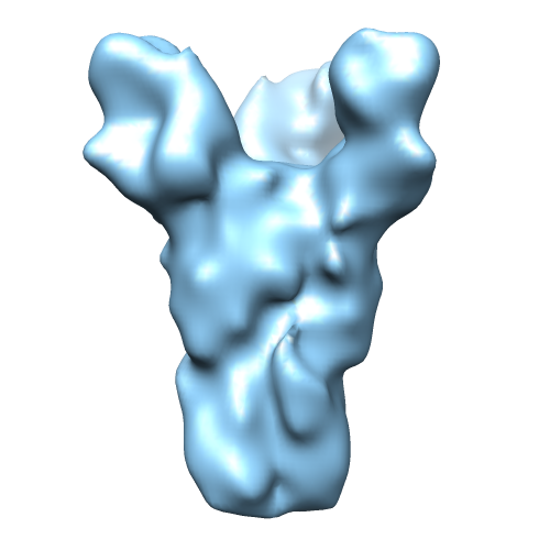
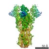
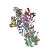
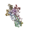
| Title | EM map of influenza H1 hemagglutatin in complex with C05 Fab | |||||||||
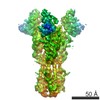
 Map data Map data | Negative stain reconstruction of H1 HA trimer in complex with three head binding C05 Fab fragments | |||||||||
 Sample Sample |
| |||||||||
 Keywords Keywords | Influenza A / flu / hemagglutanin / HA neutralizing antibody / C05 Fab | |||||||||
| Biological species |  Influenza A virus (A/Solomon Islands/3/2006(H1N1)) / Influenza A virus (A/Solomon Islands/3/2006(H1N1)) /  Homo sapiens (human) Homo sapiens (human) | |||||||||
| Method | single particle reconstruction / negative staining / Resolution: 17.0 Å | |||||||||
 Authors Authors | Khayat R / Lee JH / Ekiert DC / Wilson IA / Ward AB | |||||||||
 Citation Citation | Journal: Nature / Year: 2012 Title: Cross-neutralization of influenza A viruses mediated by a single antibody loop. Authors: Damian C Ekiert / Arun K Kashyap / John Steel / Adam Rubrum / Gira Bhabha / Reza Khayat / Jeong Hyun Lee / Michael A Dillon / Ryann E O'Neil / Aleksandr M Faynboym / Michael Horowitz / ...Authors: Damian C Ekiert / Arun K Kashyap / John Steel / Adam Rubrum / Gira Bhabha / Reza Khayat / Jeong Hyun Lee / Michael A Dillon / Ryann E O'Neil / Aleksandr M Faynboym / Michael Horowitz / Lawrence Horowitz / Andrew B Ward / Peter Palese / Richard Webby / Richard A Lerner / Ramesh R Bhatt / Ian A Wilson /  Abstract: Immune recognition of protein antigens relies on the combined interaction of multiple antibody loops, which provide a fairly large footprint and constrain the size and shape of protein surfaces that ...Immune recognition of protein antigens relies on the combined interaction of multiple antibody loops, which provide a fairly large footprint and constrain the size and shape of protein surfaces that can be targeted. Single protein loops can mediate extremely high-affinity binding, but it is unclear whether such a mechanism is available to antibodies. Here we report the isolation and characterization of an antibody called C05, which neutralizes strains from multiple subtypes of influenza A virus, including H1, H2 and H3. X-ray and electron microscopy structures show that C05 recognizes conserved elements of the receptor-binding site on the haemagglutinin surface glycoprotein. Recognition of the haemagglutinin receptor-binding site is dominated by a single heavy-chain complementarity-determining region 3 loop, with minor contacts from heavy-chain complementarity-determining region 1, and is sufficient to achieve nanomolar binding with a minimal footprint. Thus, binding predominantly with a single loop can allow antibodies to target small, conserved functional sites on otherwise hypervariable antigens. | |||||||||
| History |
|
- Structure visualization
| Movie |
 Movie viewer Movie viewer |
|---|---|
| Structure viewer | EM map: SurfViewMolmilJmol/JSmol |
| Supplemental images |
- Downloads & links
-EMDB archive
| Map data | emd_2138.map.gz | 3.5 MB | EMDB map data format | |
|---|---|---|---|---|
| Header (meta data) | emd-2138-v30.xmlemd-2138.xml | 10.7 KB 10.7 KB | Display Display | EMDB header |
| Images |  emd_2138.png emd_2138.png | 99.9 KB | ||
| Archive directory |  http://ftp.pdbj.org/pub/emdb/structures/EMD-2138ftp://ftp.pdbj.org/pub/emdb/structures/EMD-2138 http://ftp.pdbj.org/pub/emdb/structures/EMD-2138ftp://ftp.pdbj.org/pub/emdb/structures/EMD-2138 | HTTPS FTP |
-Related structure data
| Related structure data |  2139C 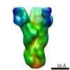 2140C 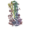 4fnkC  4fnlC  4fp8C  4fqrC C: citing same article ( |
|---|---|
| Similar structure data |

-Links
| EMDB pages | EMDB (EBI/PDBe) / EMDataResource |
|---|
-Map
| File | Download / File: emd_2138.map.gz / Format: CCP4 / Size: 3.7 MB / Type: IMAGE STORED AS FLOATING POINT NUMBER (4 BYTES) | ||||||||||||||||||||||||||||||||||||||||||||||||||||||||||||||||||||
|---|---|---|---|---|---|---|---|---|---|---|---|---|---|---|---|---|---|---|---|---|---|---|---|---|---|---|---|---|---|---|---|---|---|---|---|---|---|---|---|---|---|---|---|---|---|---|---|---|---|---|---|---|---|---|---|---|---|---|---|---|---|---|---|---|---|---|---|---|---|
| Annotation | Negative stain reconstruction of H1 HA trimer in complex with three head binding C05 Fab fragments | ||||||||||||||||||||||||||||||||||||||||||||||||||||||||||||||||||||
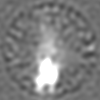
| Projections & slices | Image control
Images are generated by Spider. | ||||||||||||||||||||||||||||||||||||||||||||||||||||||||||||||||||||
| Voxel size | X=Y=Z: 2.18 Å | ||||||||||||||||||||||||||||||||||||||||||||||||||||||||||||||||||||
| Density |
| ||||||||||||||||||||||||||||||||||||||||||||||||||||||||||||||||||||
| Symmetry | Space group: 1 | ||||||||||||||||||||||||||||||||||||||||||||||||||||||||||||||||||||
| Details | EMDB XML:
CCP4 map header:
| ||||||||||||||||||||||||||||||||||||||||||||||||||||||||||||||||||||
 Z (Sec.)
 Y (Row.)
 X (Col.)


-Supplemental data
- Sample components
-Entire : Influenza HA1 from A/Solomon Islands/3/2006 in complex with C5 Fab
| Entire | Name: Influenza HA1 from A/Solomon Islands/3/2006 in complex with C5 Fab |
|---|---|
| Components |
|
-Supramolecule #1000: Influenza HA1 from A/Solomon Islands/3/2006 in complex with C5 Fab
| Supramolecule | Name: Influenza HA1 from A/Solomon Islands/3/2006 in complex with C5 Fab type: sample / ID: 1000 / Oligomeric state: One HA trimer binds to 3 fabs / Number unique components: 2 |
|---|
-Macromolecule #1: Influenza A/Solomon Islands/3/2006 hemagglutinin
| Macromolecule | Name: Influenza A/Solomon Islands/3/2006 hemagglutinin / type: protein_or_peptide / ID: 1 / Name.synonym: HA1 / Number of copies: 3 / Oligomeric state: Trimer / Recombinant expression: Yes |
|---|---|
| Source (natural) | Organism: Influenza A virus (A/Solomon Islands/3/2006(H1N1)) Strain: A/Solomon Islands/3/2006 / synonym: Flu |
| Recombinant expression | Organism: unidentified baculovirus / Recombinant plasmid: pDCE198 |
-Macromolecule #2: C05 antibody fab fragment
| Macromolecule | Name: C05 antibody fab fragment / type: protein_or_peptide / ID: 2 / Number of copies: 3 / Oligomeric state: Monomer / Recombinant expression: Yes |
|---|---|
| Source (natural) | Organism: Homo sapiens (human) / synonym: Human |
| Recombinant expression | Organism: unidentified baculovirus / Recombinant plasmid: pFastBacDual |
-Experimental details
-Structure determination
| Method | negative staining |
|---|---|
 Processing Processing | single particle reconstruction |
| Aggregation state | particle |
-Sample preparation
| Concentration | 0.1 mg/mL |
|---|---|
| Buffer | pH: 7.5 / Details: 20mM HEPES pH 7.5, 50mM NaCl |
| Staining | Type: NEGATIVE / Details: Nano-W for 30 seconds |
| Grid | Details: 400 mesh copper grid |
| Vitrification | Cryogen name: NONE / Instrument: OTHER |
- Electron microscopy
| Microscope | FEI TECNAI F20 |
|---|---|
| Temperature | Min: 293 K / Max: 293 K |
| Alignment procedure | Legacy - Astigmatism: Objective lens astigmatism corrected at 100,000x |
| Details | Images taken from 0 to 55 degrees in 5 degree tilt increments. |
| Date | Feb 14, 2012 |
| Image recording | Category: CCD / Film or detector model: GENERIC GATAN (4k x 4k) / Digitization - Sampling interval: 0.109 µm / Number real images: 252 / Average electron dose: 16 e/Å2 / Details: Images recorded using CCD / Bits/pixel: 16 |
| Tilt angle min | 0 |
| Electron beam | Acceleration voltage: 120 kV / Electron source:  FIELD EMISSION GUN FIELD EMISSION GUN |
| Electron optics | Illumination mode: FLOOD BEAM / Imaging mode: BRIGHT FIELD / Nominal defocus max: 0.9 µm / Nominal defocus min: 0.7 µm / Nominal magnification: 100000 |
| Sample stage | Specimen holder model: SIDE ENTRY, EUCENTRIC / Tilt angle max: 55 |
| Experimental equipment |  Model: Tecnai F20 / Image courtesy: FEI Company |
-Image processing
| Details | Particles were selected using an automated particle picking program. |
|---|---|
| CTF correction | Details: Micrograph and each particle |
| Final reconstruction | Applied symmetry - Point group: C3 (3 fold cyclic) / Resolution.type: BY AUTHOR / Resolution: 17.0 Å / Resolution method: FSC 0.5 CUT-OFF / Software - Name: Sparx / Number images used: 18607 |
