 Movie
Movie Controller

[English] 日本語
 Yorodumi
- EMDB-21192: human non-clustered delta protocadherin 10 on membranes tomogram 2 -
+ Open data

- Basic information
| Entry | Database: EMDB / ID: EMD-21192 | |||||||||||||||||||||
|---|---|---|---|---|---|---|---|---|---|---|---|---|---|---|---|---|---|---|---|---|---|---|
| Title | human non-clustered delta protocadherin 10 on membranes tomogram 2 | |||||||||||||||||||||
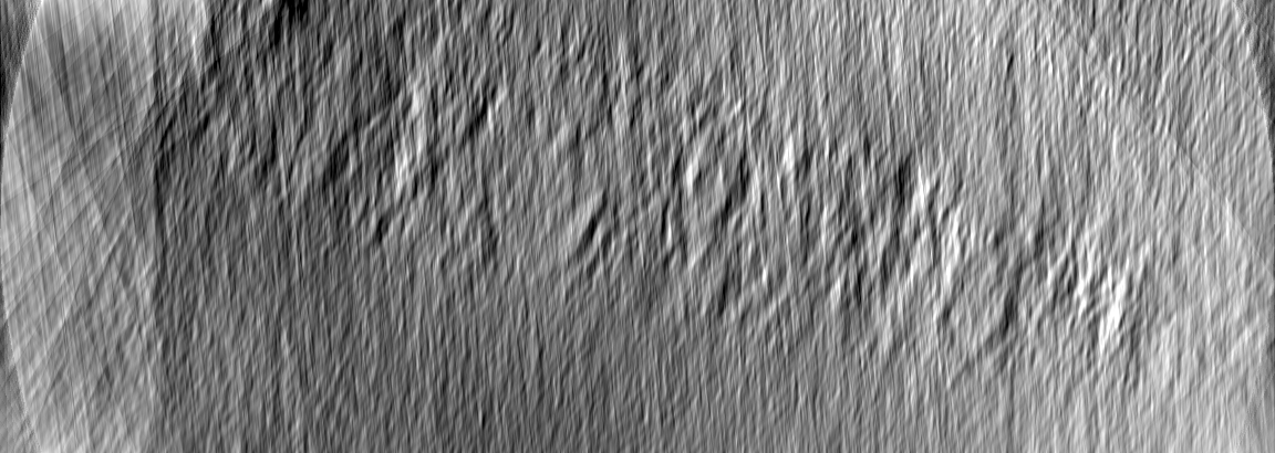
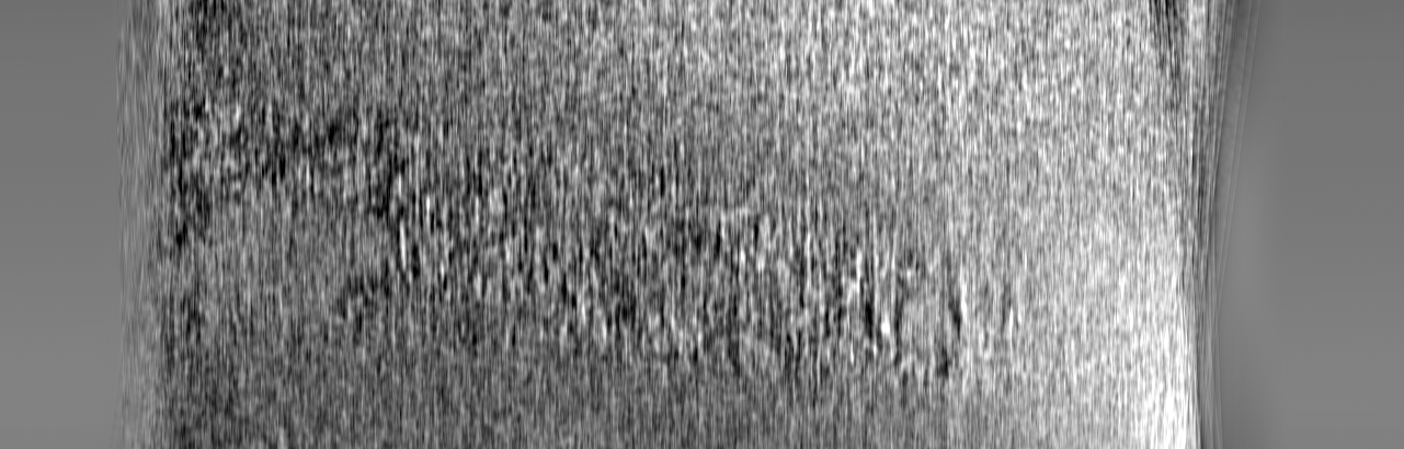
 Map data Map data | human delta protocadherin 10 full ectodomains on membranes tomogram 2 | |||||||||||||||||||||
 Sample Sample |
| |||||||||||||||||||||
| Biological species |  Homo sapiens (human) Homo sapiens (human) | |||||||||||||||||||||
| Method | electron tomography / cryo EM | |||||||||||||||||||||
 Authors Authors | Brasch J / Harrison OJ / Noble AJ / Carragher B / Potter CS | |||||||||||||||||||||
| Funding support |  United States, 6 items United States, 6 items
| |||||||||||||||||||||
 Citation Citation | Journal: Cell Rep / Year: 2020 Title: Family-wide Structural and Biophysical Analysis of Binding Interactions among Non-clustered δ-Protocadherins. Authors: Oliver J Harrison / Julia Brasch / Phinikoula S Katsamba / Goran Ahlsen / Alex J Noble / Hanbin Dan / Rosemary V Sampogna / Clinton S Potter / Bridget Carragher / Barry Honig / Lawrence Shapiro / Abstract: Non-clustered δ1- and δ2-protocadherins, close relatives of clustered protocadherins, function in cell adhesion and motility and play essential roles in neural patterning. To understand the ...Non-clustered δ1- and δ2-protocadherins, close relatives of clustered protocadherins, function in cell adhesion and motility and play essential roles in neural patterning. To understand the molecular interactions underlying these functions, we used solution biophysics to characterize binding of δ1- and δ2-protocadherins, determined crystal structures of ectodomain complexes from each family, and assessed ectodomain assembly in reconstituted intermembrane junctions by cryoelectron tomography (cryo-ET). Homophilic trans (cell-cell) interactions were preferred for all δ-protocadherins, with additional weaker heterophilic interactions observed exclusively within each subfamily. As expected, δ1- and δ2-protocadherin trans dimers formed through antiparallel EC1-EC4 interfaces, like clustered protocadherins. However, no ectodomain-mediated cis (same-cell) interactions were detectable in solution; consistent with this, cryo-ET of reconstituted junctions revealed dense assemblies lacking the characteristic order observed for clustered protocadherins. Our results define non-clustered protocadherin binding properties and their structural basis, providing a foundation for interpreting their functional roles in neural patterning. | |||||||||||||||||||||
| History |
|
- Structure visualization
| Movie |
 Movie viewer Movie viewer |
|---|---|
| Supplemental images |

- Downloads & links
-EMDB archive
| Map data | emd_21192.map.gz | 2 GB | EMDB map data format | |
|---|---|---|---|---|
| Header (meta data) | emd-21192-v30.xmlemd-21192.xml | 16.2 KB 16.2 KB | Display Display | EMDB header |
| Images | 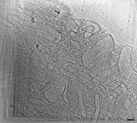 emd_21192.png emd_21192.png | 158.4 KB | ||
| Archive directory |  http://ftp.pdbj.org/pub/emdb/structures/EMD-21192ftp://ftp.pdbj.org/pub/emdb/structures/EMD-21192 http://ftp.pdbj.org/pub/emdb/structures/EMD-21192ftp://ftp.pdbj.org/pub/emdb/structures/EMD-21192 | HTTPS FTP |
-Validation report
| Summary document | emd_21192_validation.pdf.gz | 78.6 KB | Display | EMDB validaton report |
|---|---|---|---|---|
| Full document | emd_21192_full_validation.pdf.gz | 77.7 KB | Display | |
| Data in XML | emd_21192_validation.xml.gz | 499 B | Display | |
| Arichive directory | https://ftp.pdbj.org/pub/emdb/validation_reports/EMD-21192ftp://ftp.pdbj.org/pub/emdb/validation_reports/EMD-21192 | HTTPS FTP |
-Related structure data
| Related structure data |  6vfpC  6vfqC  6vfrC  6vftC  6vfuC  6vfvC  6vfwC  6vg1C  6vg4C C: citing same article ( |
|---|---|
| EM raw data | EMPIAR-10371 (Title: Human delta protocadherin 10 full ectodomains on membranes, tomogram 2 Data size: 104.6 Data #1: Appion-Protomo tilt-series alignments [tilt series]) |


-Links
| EMDB pages | EMDB (EBI/PDBe) / EMDataResource |
|---|
-Map
| File | Download / File: emd_21192.map.gz / Format: CCP4 / Size: 2.3 GB / Type: IMAGE STORED AS FLOATING POINT NUMBER (4 BYTES) | ||||||||||||||||||||||||||||||||||||||||||||||||||||||||||||||||||||
|---|---|---|---|---|---|---|---|---|---|---|---|---|---|---|---|---|---|---|---|---|---|---|---|---|---|---|---|---|---|---|---|---|---|---|---|---|---|---|---|---|---|---|---|---|---|---|---|---|---|---|---|---|---|---|---|---|---|---|---|---|---|---|---|---|---|---|---|---|---|
| Annotation | human delta protocadherin 10 full ectodomains on membranes tomogram 2 | ||||||||||||||||||||||||||||||||||||||||||||||||||||||||||||||||||||
| Projections & slices | Image control
Images are generated by Spider. generated in cubic-lattice coordinate | ||||||||||||||||||||||||||||||||||||||||||||||||||||||||||||||||||||
| Voxel size | X=Y=Z: 7.34344 Å | ||||||||||||||||||||||||||||||||||||||||||||||||||||||||||||||||||||
| Density |
| ||||||||||||||||||||||||||||||||||||||||||||||||||||||||||||||||||||
| Symmetry | Space group: 1 | ||||||||||||||||||||||||||||||||||||||||||||||||||||||||||||||||||||
| Details | EMDB XML:
CCP4 map header:
| ||||||||||||||||||||||||||||||||||||||||||||||||||||||||||||||||||||
 Z (Sec.)
 Y (Row.)
 X (Col.)


-Supplemental data
- Sample components
-Entire : trans dimers of protocadherin 10 extracellular domains formed bet...
| Entire | Name: trans dimers of protocadherin 10 extracellular domains formed between membranes of liposomes |
|---|---|
| Components |
|
-Supramolecule #1: trans dimers of protocadherin 10 extracellular domains formed bet...
| Supramolecule | Name: trans dimers of protocadherin 10 extracellular domains formed between membranes of liposomes type: complex / ID: 1 / Parent: 0 / Macromolecule list: all Details: purified full ectodomains of protocadherin 10 are tethered to Ni-NTA lipid head groups presented on the liposome surface through binding of C-terminal hexa-histidine tags |
|---|---|
| Source (natural) | Organism: Homo sapiens (human) |
-Supramolecule #2: human delta protocadherin 10
| Supramolecule | Name: human delta protocadherin 10 / type: complex / ID: 2 / Parent: 1 / Macromolecule list: all Details: full ectodomain of human delta protocadherin 10 with a C-termin hexa histidine tag |
|---|
-Supramolecule #3: liposomes
| Supramolecule | Name: liposomes / type: complex / ID: 3 / Parent: 1 Details: liposomes composed of DOPC and DOGS-NTA (8:2 ratio) with 100nm diameter |
|---|
-Macromolecule #1: human protocadherin 10
| Macromolecule | Name: human protocadherin 10 / type: protein_or_peptide / ID: 1 / Enantiomer: LEVO |
|---|---|
| Source (natural) | Organism: Homo sapiens (human) |
| Recombinant expression | Organism: Homo sapiens (human) |
| Sequence | String: SQLHYTVQEE QEHGTFVGNI AEDLGLDITK LSARGFQTVP NSR TPYLDL NLETGVLYVN EKIDREQICK QSPSCVLHLE VFLENPLELF QVEIEVLDIN DNPP SFPEP DLTVEISESA TPGTRFPLES AFDPDVGTNS LRDYEITPNS YFSLDVQTQG DGNRF AELV ...String: SQLHYTVQEE QEHGTFVGNI AEDLGLDITK LSARGFQTVP NSR TPYLDL NLETGVLYVN EKIDREQICK QSPSCVLHLE VFLENPLELF QVEIEVLDIN DNPP SFPEP DLTVEISESA TPGTRFPLES AFDPDVGTNS LRDYEITPNS YFSLDVQTQG DGNRF AELV LEKPLDREQQ AVHRYVLTAV DGGGGGGVGE GGGGGGGAGL PPQQQRTGTA LLTIRV LDS NDNVPAFDQP VYTVSLPENS PPGTLVIQLN ATDPDEGQNG EVVYSFSSHI SPRAREL FG LSPRTGRLEV SGELDYEESP VYQVYVQAKD LGPNAVPAHC KVLVRVLDAN DNAPEISF S TVKEAVSEGA APGTVVALFS VTDRDSEENG QVQCELLGDV PFRLKSSFKN YYTIVTEAP LDREAGDSYT LTVVARDRGE PALSTSKSIQ VQVSDVNDNA PRFSQPVYDV YVTENNVPGA YIYAVSATD RDEGANAQLA YSILECQIQG MSVFTYVSIN SENGYLYALR SFDYEQLKDF S FQVEARDA GSPQALAGNA TVNILIVDQN DNAPAIVAPL PGRNGTPARE VLPRSAEPGY LL TRVAAVD ADDGENARLT YSIVRGNEMN LFRMDWRTGE LRTARRVPAK RDPQRPYELV IEV RDHGQP PLSSTATLVV QLVDGAVEPQ GGGGSGGGGS GEHQRPSRSG GGETSLDLTH HHHHH |
-Experimental details
-Structure determination
| Method | cryo EM |
|---|---|
 Processing Processing | electron tomography |
| Aggregation state | 3D array |
-Sample preparation
| Concentration | 0.7 mg/mL | |||||||||||||||
|---|---|---|---|---|---|---|---|---|---|---|---|---|---|---|---|---|
| Buffer | pH: 7.4 Component:
| |||||||||||||||
| Grid | Support film - Material: CARBON / Support film - topology: LACEY / Pretreatment - Type: PLASMA CLEANING / Pretreatment - Atmosphere: OTHER / Details: unspecified | |||||||||||||||
| Vitrification | Cryogen name: ETHANE / Chamber humidity: 65 % / Chamber temperature: 298.15 K / Instrument: GATAN CRYOPLUNGE 3 / Details: 2.5 second blot before plunging.. | |||||||||||||||
| Details | aggregated liposomes were deposited onto EM grids | |||||||||||||||
| Sectioning | Other: NO SECTIONING |
- Electron microscopy
| Microscope | FEI TITAN KRIOS |
|---|---|
| Specialist optics | Phase plate: VOLTA PHASE PLATE / Spherical aberration corrector: Cs corrector (CEOS GmbH) / Energy filter - Name: GIF Bioquantum / Energy filter - Slit width: 20 eV |
| Image recording | Film or detector model: GATAN K2 QUANTUM (4k x 4k) / Detector mode: COUNTING / Number grids imaged: 1 / Number real images: 60 / Average exposure time: 1.1 sec. / Average electron dose: 3.15 e/Å2 |
| Electron beam | Acceleration voltage: 300 kV / Electron source:  FIELD EMISSION GUN FIELD EMISSION GUN |
| Electron optics | Illumination mode: FLOOD BEAM / Imaging mode: BRIGHT FIELD / Cs: 0.001 mm |
| Sample stage | Specimen holder model: FEI TITAN KRIOS AUTOGRID HOLDER / Cooling holder cryogen: NITROGEN |
| Experimental equipment |  Model: Titan Krios / Image courtesy: FEI Company |
-Image processing
| Final reconstruction | Algorithm: SIMULTANEOUS ITERATIVE (SIRT) / Software - Name: TOMO3D / Software - details: part of Appion-protomo 2.4.1 / Number images used: 53 |
|---|
