 ムービー
ムービー コントローラー


+ データを開く

- 基本情報
基本情報
| 登録情報 | データベース: EMDB / ID: EMD-21188 | |||||||||||||||||||||
|---|---|---|---|---|---|---|---|---|---|---|---|---|---|---|---|---|---|---|---|---|---|---|
| タイトル | human delta protocadherin 1 full ectodomains on membranes of liposomes tomogram 1 | |||||||||||||||||||||
 マップデータ マップデータ | human delta protocadherin 1 full ectodomains on membranes tomogram 1 | |||||||||||||||||||||
 試料 試料 |
| |||||||||||||||||||||
| 生物種 |  Homo sapiens (ヒト) Homo sapiens (ヒト) | |||||||||||||||||||||
| 手法 | 電子線トモグラフィー法 / クライオ電子顕微鏡法 | |||||||||||||||||||||
 データ登録者 データ登録者 | Brasch J / Harrisson OJ / Noble AJ / Carragher B / Potter CS / Shapiro LS | |||||||||||||||||||||
| 資金援助 |  米国, 6件 米国, 6件
| |||||||||||||||||||||
 引用 引用 | ジャーナル: Cell Rep / 年: 2020 タイトル: Family-wide Structural and Biophysical Analysis of Binding Interactions among Non-clustered δ-Protocadherins. 著者: Oliver J Harrison / Julia Brasch / Phinikoula S Katsamba / Goran Ahlsen / Alex J Noble / Hanbin Dan / Rosemary V Sampogna / Clinton S Potter / Bridget Carragher / Barry Honig / Lawrence Shapiro / 要旨: Non-clustered δ1- and δ2-protocadherins, close relatives of clustered protocadherins, function in cell adhesion and motility and play essential roles in neural patterning. To understand the ...Non-clustered δ1- and δ2-protocadherins, close relatives of clustered protocadherins, function in cell adhesion and motility and play essential roles in neural patterning. To understand the molecular interactions underlying these functions, we used solution biophysics to characterize binding of δ1- and δ2-protocadherins, determined crystal structures of ectodomain complexes from each family, and assessed ectodomain assembly in reconstituted intermembrane junctions by cryoelectron tomography (cryo-ET). Homophilic trans (cell-cell) interactions were preferred for all δ-protocadherins, with additional weaker heterophilic interactions observed exclusively within each subfamily. As expected, δ1- and δ2-protocadherin trans dimers formed through antiparallel EC1-EC4 interfaces, like clustered protocadherins. However, no ectodomain-mediated cis (same-cell) interactions were detectable in solution; consistent with this, cryo-ET of reconstituted junctions revealed dense assemblies lacking the characteristic order observed for clustered protocadherins. Our results define non-clustered protocadherin binding properties and their structural basis, providing a foundation for interpreting their functional roles in neural patterning. | |||||||||||||||||||||
| 履歴 |
|
- 構造の表示
構造の表示
| ムービー |
 ムービービューア ムービービューア |
|---|---|
| 添付画像 |

- ダウンロードとリンク
-EMDBアーカイブ
| マップデータ | emd_21188.map.gz | 1.5 GB | EMDBマップデータ形式 | |
|---|---|---|---|---|
| ヘッダ (付随情報) | emd-21188-v30.xmlemd-21188.xml | 16.3 KB 16.3 KB | 表示 表示 | EMDBヘッダ |
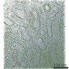
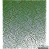
| 画像 | 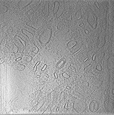 emd_21188.png emd_21188.png | 138.5 KB | ||
| アーカイブディレクトリ |  http://ftp.pdbj.org/pub/emdb/structures/EMD-21188ftp://ftp.pdbj.org/pub/emdb/structures/EMD-21188 http://ftp.pdbj.org/pub/emdb/structures/EMD-21188ftp://ftp.pdbj.org/pub/emdb/structures/EMD-21188 | HTTPS FTP |
-検証レポート
| 文書・要旨 | emd_21188_validation.pdf.gz | 78.4 KB | 表示 | EMDB検証レポート |
|---|---|---|---|---|
| 文書・詳細版 | emd_21188_full_validation.pdf.gz | 77.4 KB | 表示 | |
| XML形式データ | emd_21188_validation.xml.gz | 499 B | 表示 | |
| アーカイブディレクトリ | https://ftp.pdbj.org/pub/emdb/validation_reports/EMD-21188ftp://ftp.pdbj.org/pub/emdb/validation_reports/EMD-21188 | HTTPS FTP |
-関連構造データ
| 関連構造データ |  6vfpC  6vfqC  6vfrC  6vftC  6vfuC  6vfvC  6vfwC  6vg1C  6vg4C C: 同じ文献を引用 ( |
|---|---|
| 電子顕微鏡画像生データ | EMPIAR-10367 (タイトル: Human delta protocadherin 1 full ectodomains on membranes, tomogram 1 Data size: 247.7 Data #1: Summed, non-frame aligned tilt images [micrographs - single frame] Data #2: Appion-Protomo tilt-series alignments [tilt series]) |


-リンク
| EMDBのページ | EMDB (EBI/PDBe) / EMDataResource |
|---|
-マップ
| ファイル | ダウンロード / ファイル: emd_21188.map.gz / 形式: CCP4 / 大きさ: 1.7 GB / タイプ: IMAGE STORED AS FLOATING POINT NUMBER (4 BYTES) | ||||||||||||||||||||||||||||||||||||||||||||||||||||||||||||||||||||
|---|---|---|---|---|---|---|---|---|---|---|---|---|---|---|---|---|---|---|---|---|---|---|---|---|---|---|---|---|---|---|---|---|---|---|---|---|---|---|---|---|---|---|---|---|---|---|---|---|---|---|---|---|---|---|---|---|---|---|---|---|---|---|---|---|---|---|---|---|---|
| 注釈 | human delta protocadherin 1 full ectodomains on membranes tomogram 1 | ||||||||||||||||||||||||||||||||||||||||||||||||||||||||||||||||||||
| 投影像・断面図 | 画像のコントロール
画像は Spider により作成 これらの図は立方格子座標系で作成されたものです | ||||||||||||||||||||||||||||||||||||||||||||||||||||||||||||||||||||
| ボクセルのサイズ | X=Y=Z: 9.19 Å | ||||||||||||||||||||||||||||||||||||||||||||||||||||||||||||||||||||
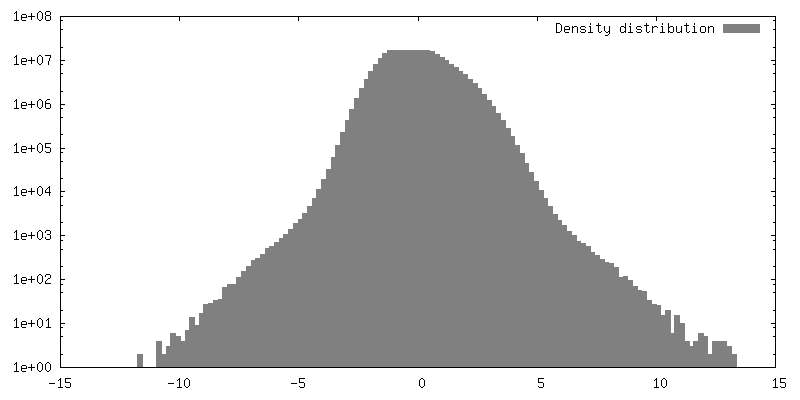
| 密度 |
| ||||||||||||||||||||||||||||||||||||||||||||||||||||||||||||||||||||
| 対称性 | 空間群: 1 | ||||||||||||||||||||||||||||||||||||||||||||||||||||||||||||||||||||
| 詳細 | EMDB XML:
CCP4マップ ヘッダ情報:
| ||||||||||||||||||||||||||||||||||||||||||||||||||||||||||||||||||||
 Z (Sec.)
 Y (Row.)
 X (Col.)


-添付データ
- 試料の構成要素
-全体 : trans dimers of protocadherin 1 extracellular domains formed betw...
| 全体 | 名称: trans dimers of protocadherin 1 extracellular domains formed between membranes of liposomes |
|---|---|
| 要素 |
|
-超分子 #1: trans dimers of protocadherin 1 extracellular domains formed betw...
| 超分子 | 名称: trans dimers of protocadherin 1 extracellular domains formed between membranes of liposomes タイプ: complex / ID: 1 / 親要素: 0 / 含まれる分子: all 詳細: purified full ectodomains of protocadherin 1 are tethered to Ni-NTA lipid head groups presented on the liposome surface through binding of C-terminal hexa-histidine tags |
|---|---|
| 由来(天然) | 生物種: Homo sapiens (ヒト) |
-超分子 #2: human delta protocadherin 1
| 超分子 | 名称: human delta protocadherin 1 / タイプ: complex / ID: 2 / 親要素: 1 / 含まれる分子: all 詳細: full ectodomain of human delta protocadherin 1 with a C-termin hexa histidine tag |
|---|
-超分子 #3: liposomes
| 超分子 | 名称: liposomes / タイプ: complex / ID: 3 / 親要素: 1 詳細: liposomes composed of DOPC and DOGS-NTA (8:2 ratio) with 100nm diameter |
|---|
-分子 #1: human delta protocadherin 1
| 分子 | 名称: human delta protocadherin 1 / タイプ: protein_or_peptide / ID: 1 詳細: BiP-signal sequence and hexa histidine C-terminal tag. 光学異性体: LEVO |
|---|---|
| 由来(天然) | 生物種: Homo sapiens (ヒト) |
| 組換発現 | 生物種: Homo sapiens (ヒト) |
| 配列 | 文字列: MKLSLVAAML LLLSAARATR VVYKVPEEQP PNTLIGSLAA DYGFPDVGHL YKLEVGAPYL RVDGKTGDIF TTETSIDR E GLRECQNQLP GDPCILEFEV SITDLVQNGS PRLLEGQIEV QDINDNTPNF ASPVITLAI PENTNIGSLF PIPLASDRDA GPNGVASYEL ...文字列: MKLSLVAAML LLLSAARATR VVYKVPEEQP PNTLIGSLAA DYGFPDVGHL YKLEVGAPYL RVDGKTGDIF TTETSIDR E GLRECQNQLP GDPCILEFEV SITDLVQNGS PRLLEGQIEV QDINDNTPNF ASPVITLAI PENTNIGSLF PIPLASDRDA GPNGVASYEL QAGPEAQELF GLQVAEDQEE KQPQLIVMGN LDRERWDSY DLTIKVQDGG SPPRASSALL RVTVLDTNDN APKFERPSYE AELSENSPIG H SVIQVKAN DSDQGANAEI EYTFHQAPEV VRRLLRLDRN TGLITVQGPV DREDLSTLRF SV LAKDRGT NPKSARAQVV VTVKDMNDNA PTIEIRGIGL VTHQDGMANI SEDVAEETAV ALV QVSDRD EGENAAVTCV VAGDVPFQLR QASETGSDSK KKYFLQTTTP LDYEKVKDYT IEIV AVDSG NPPLSSTNSL KVQVVDVNDN APVFTQSVTE VAFPENNKPG EVIAEITASD ADSGS NAEL VYSLEPEPAA KGLFTISPET GEIQVKTSLD REQRESYELK VVAADRGSPS LQGTAT VLV NVLDCNDNDP KFMLSGYNFS VMENMPALSP VGMVTVIDGD KGENAQVQLS VEQDNGD FV IQNGTGTILS SLSFDREQQS TYTFQLKAVD GGVPPRSAYV GVTINVLDEN DNAPYITA P SNTSHKLLTP QTRLGETVSQ VAAEDFDSGV NAELIYSIAG GNPYGLFQIG SHSGAITLE KEIERRHHGL HRLVVKVSDR GKPPRYGTAL VHLYVNETLA NRTLLETLLG HSLDTPLDID IAGDPEYER SKQRGNHHHH HH |
-実験情報
-構造解析
| 手法 | クライオ電子顕微鏡法 |
|---|---|
 解析 解析 | 電子線トモグラフィー法 |
| 試料の集合状態 | 3D array |
-試料調製
| 濃度 | 0.7 mg/mL | |||||||||||||||
|---|---|---|---|---|---|---|---|---|---|---|---|---|---|---|---|---|
| 緩衝液 | pH: 7.4 構成要素:
| |||||||||||||||
| グリッド | 支持フィルム - 材質: CARBON / 支持フィルム - トポロジー: LACEY / 詳細: unspecified | |||||||||||||||
| 凍結 | 凍結剤: ETHANE / チャンバー内湿度: 65 % / チャンバー内温度: 298.15 K / 装置: GATAN CRYOPLUNGE 3 / 詳細: 2.5 second blot before plunging.. | |||||||||||||||
| 詳細 | aggregated liposomes were deposited onto EM grids. | |||||||||||||||
| 切片作成 | その他: NO SECTIONING |
- 電子顕微鏡法
| 顕微鏡 | FEI TITAN KRIOS |
|---|---|
| 特殊光学系 | 位相板: VOLTA PHASE PLATE / 球面収差補正装置: Cs corrector (CEOS GmbH) / エネルギーフィルター - 名称: GIF Bioquantum / エネルギーフィルター - スリット幅: 20 eV |
| 撮影 | フィルム・検出器のモデル: GATAN K2 QUANTUM (4k x 4k) 検出モード: COUNTING / 撮影したグリッド数: 1 / 実像数: 47 / 平均露光時間: 1.52 sec. / 平均電子線量: 1.72 e/Å2 |
| 電子線 | 加速電圧: 300 kV / 電子線源:  FIELD EMISSION GUN FIELD EMISSION GUN |
| 電子光学系 | 照射モード: FLOOD BEAM / 撮影モード: BRIGHT FIELD / Cs: 0.001 mm |
| 試料ステージ | 試料ホルダーモデル: FEI TITAN KRIOS AUTOGRID HOLDER ホルダー冷却材: NITROGEN |
| 実験機器 |  モデル: Titan Krios / 画像提供: FEI Company |
-画像解析
| 最終 再構成 | アルゴリズム: SIMULTANEOUS ITERATIVE (SIRT) / ソフトウェア - 名称: TOMO3D ソフトウェア - 詳細: part of Appion-protomo 2.4.1 TOMO3D SIRT 使用した粒子像数: 38 |
|---|
