 Movie
Movie Controller


+ Open data

- Basic information
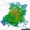
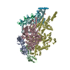
| Entry | Database: EMDB / ID: EMD-21102 | |||||||||
|---|---|---|---|---|---|---|---|---|---|---|
| Title | RSC body (classified) | |||||||||
 Map data Map data | RSC body (classified) | |||||||||
 Sample Sample |
| |||||||||
| Biological species |  | |||||||||
| Method | single particle reconstruction / cryo EM / Resolution: 3.34 Å | |||||||||
 Authors Authors | Patel AB / Moore CM / Greber BJ / Nogales E | |||||||||
| Funding support |  United States, 2 items United States, 2 items
| |||||||||
 Citation Citation | Journal: Elife / Year: 2019 Title: Architecture of the chromatin remodeler RSC and insights into its nucleosome engagement. Authors: Avinash B Patel / Camille M Moore / Basil J Greber / Jie Luo / Stefan A Zukin / Jeff Ranish / Eva Nogales / Abstract: Eukaryotic DNA is packaged into nucleosome arrays, which are repositioned by chromatin remodeling complexes to control DNA accessibility. The RSC (emodeling the tructure of hromatin) complex, a ...Eukaryotic DNA is packaged into nucleosome arrays, which are repositioned by chromatin remodeling complexes to control DNA accessibility. The RSC (emodeling the tructure of hromatin) complex, a member of the SWI/SNF chromatin remodeler family, plays critical roles in genome maintenance, transcription, and DNA repair. Here, we report cryo-electron microscopy (cryo-EM) and crosslinking mass spectrometry (CLMS) studies of yeast RSC complex and show that RSC is composed of a rigid tripartite core and two flexible lobes. The core structure is scaffolded by an asymmetric Rsc8 dimer and built with the evolutionarily conserved subunits Sfh1, Rsc6, Rsc9 and Sth1. The flexible ATPase lobe, composed of helicase subunit Sth1, Arp7, Arp9 and Rtt102, is anchored to this core by the N-terminus of Sth1. Our cryo-EM analysis of RSC bound to a nucleosome core particle shows that in addition to the expected nucleosome-Sth1 interactions, RSC engages histones and nucleosomal DNA through one arm of the core structure, composed of the Rsc8 SWIRM domains, Sfh1 and Npl6. Our findings provide structural insights into the conserved assembly process for all members of the SWI/SNF family of remodelers, and illustrate how RSC selects, engages, and remodels nucleosomes. | |||||||||
| History |
|
- Structure visualization
| Movie |
 Movie viewer Movie viewer |
|---|---|
| Structure viewer | EM map: SurfViewMolmilJmol/JSmol |
| Supplemental images |

- Downloads & links
-EMDB archive
| Map data | emd_21102.map.gz | 85 MB | EMDB map data format | |
|---|---|---|---|---|
| Header (meta data) | emd-21102-v30.xmlemd-21102.xml | 15.4 KB 15.4 KB | Display Display | EMDB header |
| FSC (resolution estimation) | emd_21102_fsc.xml | 10.2 KB | Display | FSC data file |
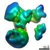
| Images | 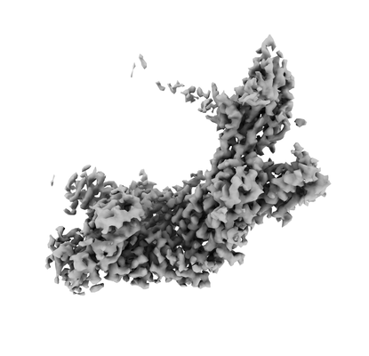 emd_21102.png emd_21102.png | 43.5 KB | ||
| Masks | emd_21102_msk_1.map | 91.1 MB | Mask map | |
| Others | emd_21102_half_map_1.map.gzemd_21102_half_map_2.map.gz | 71.3 MB 71.2 MB | ||
| Archive directory |  http://ftp.pdbj.org/pub/emdb/structures/EMD-21102ftp://ftp.pdbj.org/pub/emdb/structures/EMD-21102 http://ftp.pdbj.org/pub/emdb/structures/EMD-21102ftp://ftp.pdbj.org/pub/emdb/structures/EMD-21102 | HTTPS FTP |
-Related structure data


-Links
| EMDB pages | EMDB (EBI/PDBe) / EMDataResource |
|---|
-Map
| File | Download / File: emd_21102.map.gz / Format: CCP4 / Size: 91.1 MB / Type: IMAGE STORED AS FLOATING POINT NUMBER (4 BYTES) | ||||||||||||||||||||||||||||||||||||||||||||||||||||||||||||
|---|---|---|---|---|---|---|---|---|---|---|---|---|---|---|---|---|---|---|---|---|---|---|---|---|---|---|---|---|---|---|---|---|---|---|---|---|---|---|---|---|---|---|---|---|---|---|---|---|---|---|---|---|---|---|---|---|---|---|---|---|---|
| Annotation | RSC body (classified) | ||||||||||||||||||||||||||||||||||||||||||||||||||||||||||||
| Projections & slices | Image control
Images are generated by Spider. | ||||||||||||||||||||||||||||||||||||||||||||||||||||||||||||
| Voxel size | X=Y=Z: 1.439 Å | ||||||||||||||||||||||||||||||||||||||||||||||||||||||||||||
| Density |
| ||||||||||||||||||||||||||||||||||||||||||||||||||||||||||||
| Symmetry | Space group: 1 | ||||||||||||||||||||||||||||||||||||||||||||||||||||||||||||
| Details | EMDB XML:
CCP4 map header:
| ||||||||||||||||||||||||||||||||||||||||||||||||||||||||||||
 Z (Sec.)
 Y (Row.)
 X (Col.)


-Supplemental data
-Mask #1
| File | emd_21102_msk_1.map | ||||||||||||
|---|---|---|---|---|---|---|---|---|---|---|---|---|---|
| Projections & Slices |
| ||||||||||||
| Density Histograms |


-Half map: RSC body (classified)
| File | emd_21102_half_map_1.map | ||||||||||||
|---|---|---|---|---|---|---|---|---|---|---|---|---|---|
| Annotation | RSC body (classified) | ||||||||||||
| Projections & Slices |
| ||||||||||||
| Density Histograms |


-Half map: RSC body (classified)
| File | emd_21102_half_map_2.map | ||||||||||||
|---|---|---|---|---|---|---|---|---|---|---|---|---|---|
| Annotation | RSC body (classified) | ||||||||||||
| Projections & Slices |
| ||||||||||||
| Density Histograms |


- Sample components
-Entire : Remodeling the Structure of Chromatin (RSC)
| Entire | Name: Remodeling the Structure of Chromatin (RSC) |
|---|---|
| Components |
|
-Supramolecule #1: Remodeling the Structure of Chromatin (RSC)
| Supramolecule | Name: Remodeling the Structure of Chromatin (RSC) / type: complex / ID: 1 / Parent: 0 |
|---|---|
| Source (natural) | Organism: |
| Molecular weight | Theoretical: 1.1 MDa |
-Experimental details
-Structure determination
| Method | cryo EM |
|---|---|
 Processing Processing | single particle reconstruction |
| Aggregation state | particle |
-Sample preparation
| Concentration | .5 mg/mL |
|---|---|
| Buffer | pH: 7.9 |
| Grid | Support film - Material: CARBON / Support film - topology: HOLEY ARRAY / Details: unspecified |
| Vitrification | Cryogen name: ETHANE / Chamber humidity: 100 % / Chamber temperature: 277.15 K / Instrument: FEI VITROBOT MARK IV / Details: BT 2-3s; BF 0N. |
- Electron microscopy
| Microscope | FEI TITAN KRIOS |
|---|---|
| Specialist optics | Energy filter - Name: GIF Bioquantum / Energy filter - Slit width: 20 eV |
| Image recording | Film or detector model: GATAN K3 BIOQUANTUM (6k x 4k) / Number grids imaged: 3 / Number real images: 8122 / Average exposure time: 3.0 sec. / Average electron dose: 40.0 e/Å2 |
| Electron beam | Acceleration voltage: 300 kV / Electron source:  FIELD EMISSION GUN FIELD EMISSION GUN |
| Electron optics | C2 aperture diameter: 50.0 µm / Calibrated magnification: 46339 / Illumination mode: FLOOD BEAM / Imaging mode: BRIGHT FIELD / Cs: 2.7 mm / Nominal defocus min: 1.0 µm |
| Sample stage | Specimen holder model: FEI TITAN KRIOS AUTOGRID HOLDER / Cooling holder cryogen: NITROGEN |
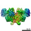
| Experimental equipment |  Model: Titan Krios / Image courtesy: FEI Company |
-Image processing
| Final reconstruction | Resolution.type: BY AUTHOR / Resolution: 3.34 Å / Resolution method: FSC 0.143 CUT-OFF / Number images used: 43905 |
|---|---|
| Initial angle assignment | Type: MAXIMUM LIKELIHOOD |
| Final angle assignment | Type: MAXIMUM LIKELIHOOD |
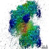
| FSC plot (resolution estimation) |  |
