 Movie
Movie Controller

[English] 日本語
 Yorodumi
- EMDB-21097: Negative stain electron microscopy of PmPV2, a toxin member of th... -
+ Open data

- Basic information
| Entry | Database: EMDB / ID: EMD-21097 | ||||||||||||
|---|---|---|---|---|---|---|---|---|---|---|---|---|---|
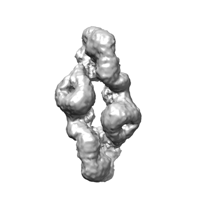
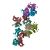
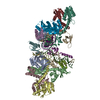
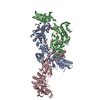
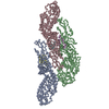
| Title | Negative stain electron microscopy of PmPV2, a toxin member of the Membrane Attack Complex-Perforin (MACPF) from the eggs of Pomacea maculata | ||||||||||||
 Map data Map data | PmPV2 | ||||||||||||
 Sample Sample |
| ||||||||||||
 Keywords Keywords | Perivitellin-2 / Pomacea maculata / PmPV2 / Apple snail / Gastropod reproduction / Anti-predator defense. / TOXIN | ||||||||||||
| Biological species |  Pomacea maculata (invertebrata) Pomacea maculata (invertebrata) | ||||||||||||
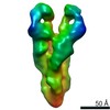
| Method | single particle reconstruction / negative staining / Resolution: 15.2 Å | ||||||||||||
 Authors Authors | Otero LH / Brola TR / Giglio ML / Ituarte S / Dreon MS / Heras H | ||||||||||||
| Funding support |  Argentina, 3 items Argentina, 3 items
| ||||||||||||
 Citation Citation | Journal: J Struct Biol / Year: 2020 Title: Exaptation of two ancient immune proteins into a new dimeric pore-forming toxin in snails. Authors: M L Giglio / S Ituarte / V Milesi / M S Dreon / T R Brola / J Caramelo / J C H Ip / S Maté / J W Qiu / L H Otero / H Heras /  Abstract: The Membrane Attack Complex-Perforin (MACPF) family is ubiquitously found in all kingdoms. They have diverse cellular roles, however MACPFs with pore-forming toxic function in venoms and poisons are ...The Membrane Attack Complex-Perforin (MACPF) family is ubiquitously found in all kingdoms. They have diverse cellular roles, however MACPFs with pore-forming toxic function in venoms and poisons are very rare in animals. Here we present the structure of PmPV2, a MACPF toxin from the poisonous apple snail eggs, that can affect the digestive and nervous systems of potential predators. We report the three-dimensional structure of PmPV2, at 17.2 Å resolution determined by negative-stain electron microscopy and its solution structure by small angle X-ray scattering (SAXS). We found that PV2s differ from nearly all MACPFs in two respects: it is a dimer in solution and protomers combine two immune proteins into an AB toxin. The MACPF chain is linked by a single disulfide bond to a tachylectin chain, and two heterodimers are arranged head-to-tail by non-covalent forces in the native protein. MACPF domain is fused with a putative new Ct-accessory domain exclusive to invertebrates. The tachylectin is a six-bladed β-propeller, similar to animal tectonins. We experimentally validated the predicted functions of both subunits and demonstrated for the first time that PV2s are true pore-forming toxins. The tachylectin "B" delivery subunit would bind to target membranes, and then the MACPF "A" toxic subunit would disrupt lipid bilayers forming large pores altering the plasma membrane conductance. These results indicate that PV2s toxicity evolved by linking two immune proteins where their combined preexisting functions gave rise to a new toxic entity with a novel role in defense against predation. This structure is an unparalleled example of protein exaptation. | ||||||||||||
| History |
|
- Structure visualization
| Movie |
 Movie viewer Movie viewer |
|---|---|
| Structure viewer | EM map: SurfViewMolmilJmol/JSmol |
| Supplemental images |
- Downloads & links
-EMDB archive
| Map data | emd_21097.map.gz | 25 MB | EMDB map data format | |
|---|---|---|---|---|
| Header (meta data) | emd-21097-v30.xmlemd-21097.xml | 15.3 KB 15.3 KB | Display Display | EMDB header |
| Images |  emd_21097.png emd_21097.png | 31.8 KB | ||
| Filedesc metadata | emd-21097.cif.gz | 5.5 KB | ||
| Archive directory |  http://ftp.pdbj.org/pub/emdb/structures/EMD-21097ftp://ftp.pdbj.org/pub/emdb/structures/EMD-21097 http://ftp.pdbj.org/pub/emdb/structures/EMD-21097ftp://ftp.pdbj.org/pub/emdb/structures/EMD-21097 | HTTPS FTP |
-Validation report
| Summary document | emd_21097_validation.pdf.gz | 530.7 KB | Display | EMDB validaton report |
|---|---|---|---|---|
| Full document | emd_21097_full_validation.pdf.gz | 530.2 KB | Display | |
| Data in XML | emd_21097_validation.xml.gz | 6 KB | Display | |
| Data in CIF | emd_21097_validation.cif.gz | 6.8 KB | Display | |
| Arichive directory | https://ftp.pdbj.org/pub/emdb/validation_reports/EMD-21097ftp://ftp.pdbj.org/pub/emdb/validation_reports/EMD-21097 | HTTPS FTP |
-Related structure data
| Similar structure data |
|---|


-Links
| EMDB pages | EMDB (EBI/PDBe) / EMDataResource |
|---|
-Map
| File | Download / File: emd_21097.map.gz / Format: CCP4 / Size: 27 MB / Type: IMAGE STORED AS FLOATING POINT NUMBER (4 BYTES) | ||||||||||||||||||||||||||||||||||||||||||||||||||||||||||||||||||||
|---|---|---|---|---|---|---|---|---|---|---|---|---|---|---|---|---|---|---|---|---|---|---|---|---|---|---|---|---|---|---|---|---|---|---|---|---|---|---|---|---|---|---|---|---|---|---|---|---|---|---|---|---|---|---|---|---|---|---|---|---|---|---|---|---|---|---|---|---|---|
| Annotation | PmPV2 | ||||||||||||||||||||||||||||||||||||||||||||||||||||||||||||||||||||
| Voxel size | X=Y=Z: 2.02 Å | ||||||||||||||||||||||||||||||||||||||||||||||||||||||||||||||||||||
| Density |
| ||||||||||||||||||||||||||||||||||||||||||||||||||||||||||||||||||||
| Symmetry | Space group: 1 | ||||||||||||||||||||||||||||||||||||||||||||||||||||||||||||||||||||
| Details | EMDB XML:
CCP4 map header:
| ||||||||||||||||||||||||||||||||||||||||||||||||||||||||||||||||||||
-Supplemental data
- Sample components
-Entire : Perivitellin PmPV2
| Entire | Name: Perivitellin PmPV2 |
|---|---|
| Components |
|
-Supramolecule #1: Perivitellin PmPV2
| Supramolecule | Name: Perivitellin PmPV2 / type: complex / ID: 1 / Parent: 0 / Macromolecule list: all Details: Protein from the Pomacea maculata eggs. Its structure consists of a dimer of heterodimer, each consisting in a Tachylectin (PmPV2-31) and a MACPF-containing protein (PmPV2-67) held together by a disulfide bond. |
|---|---|
| Source (natural) | Organism: Pomacea maculata (invertebrata) |
| Molecular weight | Theoretical: 162 KDa |
-Macromolecule #1: Tachylectin subunit (PmPV2-31)
| Macromolecule | Name: Tachylectin subunit (PmPV2-31) / type: protein_or_peptide / ID: 1 / Details: Protein without signal peptide / Enantiomer: LEVO |
|---|---|
| Source (natural) | Organism: Pomacea maculata (invertebrata) |
| Sequence | String: FTSVKLPRDE HWPYNYVSVG PAGVWAVNRQ NKLFYRTGTY GDNANMGSGW QFKQDGVGQV DVGKDKVGY INLSGGSLFR IEGISQANPV GGTPKSWEWW TKYIGMSLRE DTRFSSRIEN Q NKVLTFTF RTCFWASRIT NWCFADSSYT ETVTAGGSGT WITKSQLKYK ...String: FTSVKLPRDE HWPYNYVSVG PAGVWAVNRQ NKLFYRTGTY GDNANMGSGW QFKQDGVGQV DVGKDKVGY INLSGGSLFR IEGISQANPV GGTPKSWEWW TKYIGMSLRE DTRFSSRIEN Q NKVLTFTF RTCFWASRIT NWCFADSSYT ETVTAGGSGT WITKSQLKYK SGTFGNPDTE GG DWILVDS GSFQHVSSGS GVVLAVRSNG ELVQRTGITC SLPQGTGWTS MLNSMSRVDT YGT VAWAVD TAGDLYFINL |
-Macromolecule #2: MACPF subunit (PmPV2-67)
| Macromolecule | Name: MACPF subunit (PmPV2-67) / type: protein_or_peptide / ID: 2 / Enantiomer: LEVO |
|---|---|
| Source (natural) | Organism: Pomacea maculata (invertebrata) |
| Sequence | String: ARVCPKIVPG LDKLRVGVDI TKLDLLPLFD LGDNGFRSAV ADYTCDRGQT AVVDGESFDV PDQVDSVVI ESSGQQTSSV TTIKSESQIS QALSISAGIS VETAKAGFSS SASYAEMQEA I TKYGRTVS QMSAVYTTCS ANLSPNLLLG QNPLQTLSRL PSDFTADTQG ...String: ARVCPKIVPG LDKLRVGVDI TKLDLLPLFD LGDNGFRSAV ADYTCDRGQT AVVDGESFDV PDQVDSVVI ESSGQQTSSV TTIKSESQIS QALSISAGIS VETAKAGFSS SASYAEMQEA I TKYGRTVS QMSAVYTTCS ANLSPNLLLG QNPLQTLSRL PSDFTADTQG YYDFIKTYGT HY FNKGKLG GMFLFTSETD MSYFQNKNSQ QIEATVKATF ASILSTETGG SSDESKEVIE FKE SSLITS KFFGGQTNLA ADGLTKWQPT IAKLPYFMSG TLSTISSLIA DTTKRASMEL AVKN YLLKA KVANLDRLTY IRLNSWSVGH NELRDLSAQL QNLKTKTIFS DADEKLLQSI EDQVS VPAW FSDRTTFCFR STAVGSADQC NGQSTNTLCA EPNRYTQQYM DKTYLGDTGC RLVWKI STT ESTDWFKSVK VNFRWYPTWS PCACGPVGTP FTISAPANSW TQDYLDVTNP KFGECML QW MIEVPPTATL WAKNLEFCID FTCGKKKQCV DANQWTEPYL DISAHEACGM SWALIAK |
-Experimental details
-Structure determination
| Method | negative staining |
|---|---|
 Processing Processing | single particle reconstruction |
| Aggregation state | particle |
-Sample preparation
| Concentration | 0.05 mg/mL | ||||||
|---|---|---|---|---|---|---|---|
| Buffer | pH: 8.5 / Component:
| ||||||
| Staining | Type: NEGATIVE / Material: Uranyl Acetate | ||||||
| Grid | Material: COPPER / Mesh: 400 / Support film - Material: CARBON / Support film - topology: LACEY / Pretreatment - Type: GLOW DISCHARGE / Pretreatment - Time: 50 sec. / Pretreatment - Atmosphere: AIR / Pretreatment - Pressure: 0.037 kPa / Details: Negative glow discharge 25 mA | ||||||
| Details | The sample was monodisperse |
- Electron microscopy
| Microscope | FEI TALOS ARCTICA |
|---|---|
| Image recording | Film or detector model: FEI CETA (4k x 4k) / Digitization - Dimensions - Width: 4096 pixel / Digitization - Dimensions - Height: 4096 pixel / Number grids imaged: 1 / Number real images: 60 / Average exposure time: 1.0 sec. / Average electron dose: 20.0 e/Å2 / Details: About 400 particles per image |
| Electron beam | Acceleration voltage: 200 kV / Electron source:  FIELD EMISSION GUN FIELD EMISSION GUN |
| Electron optics | C2 aperture diameter: 100.0 µm / Calibrated defocus max: 4.0 µm / Calibrated defocus min: 2.0 µm / Calibrated magnification: 73000 / Illumination mode: OTHER / Imaging mode: BRIGHT FIELD / Cs: 2.7 mm / Nominal defocus max: 4.0 µm / Nominal defocus min: 2.0 µm / Nominal magnification: 73000 |
| Sample stage | Specimen holder model: OTHER |
| Experimental equipment |  Model: Talos Arctica / Image courtesy: FEI Company |
