 Movie
Movie Controller

[English] 日本語
 Yorodumi
- EMDB-20759: Structure of recombinantly assembled E46K alpha-synuclein fibrils -
+ Open data

- Basic information
| Entry | Database: EMDB / ID: EMD-20759 | ||||||||||||
|---|---|---|---|---|---|---|---|---|---|---|---|---|---|
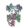
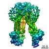
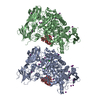
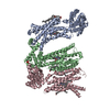
| Title | Structure of recombinantly assembled E46K alpha-synuclein fibrils | ||||||||||||
 Map data Map data | |||||||||||||
 Sample Sample |
| ||||||||||||
 Keywords Keywords | alpha-synuclein / amyloid / fibril / E46K / hereditary mutations / parkinson's disease / lewy body dementia / PROTEIN FIBRIL | ||||||||||||
| Function / homology |  Function and homology information Function and homology informationnegative regulation of mitochondrial electron transport, NADH to ubiquinone / neutral lipid metabolic process / regulation of acyl-CoA biosynthetic process / negative regulation of dopamine uptake involved in synaptic transmission / negative regulation of norepinephrine uptake / response to desipramine / positive regulation of SNARE complex assembly / positive regulation of hydrogen peroxide catabolic process / supramolecular fiber / regulation of synaptic vesicle recycling ...negative regulation of mitochondrial electron transport, NADH to ubiquinone / neutral lipid metabolic process / regulation of acyl-CoA biosynthetic process / negative regulation of dopamine uptake involved in synaptic transmission / negative regulation of norepinephrine uptake / response to desipramine / positive regulation of SNARE complex assembly / positive regulation of hydrogen peroxide catabolic process / supramolecular fiber / regulation of synaptic vesicle recycling / negative regulation of chaperone-mediated autophagy / regulation of reactive oxygen species biosynthetic process / positive regulation of protein localization to cell periphery / mitochondrial membrane organization / negative regulation of exocytosis / regulation of glutamate secretion / dopamine biosynthetic process / regulation of macrophage activation / positive regulation of neurotransmitter secretion / response to iron(II) ion / negative regulation of dopamine metabolic process / negative regulation of platelet-derived growth factor receptor signaling pathway / SNARE complex assembly / negative regulation of thrombin-activated receptor signaling pathway / Lewy body / regulation of locomotion / negative regulation of microtubule polymerization / synaptic vesicle priming / regulation of norepinephrine uptake / transporter regulator activity / protein kinase inhibitor activity / positive regulation of inositol phosphate biosynthetic process / synaptic vesicle transport / dopamine uptake involved in synaptic transmission / regulation of dopamine secretion / positive regulation of receptor recycling / cuprous ion binding / positive regulation of exocytosis / nuclear outer membrane / mitochondrial ATP synthesis coupled electron transport / dynein complex binding / synaptic vesicle exocytosis / response to magnesium ion / positive regulation of endocytosis / negative regulation of serotonin uptake / response to type II interferon / cysteine-type endopeptidase inhibitor activity / kinesin binding / regulation of presynapse assembly / synaptic vesicle endocytosis / alpha-tubulin binding / beta-tubulin binding / phospholipase binding / phospholipid metabolic process / supramolecular fiber organization / behavioral response to cocaine / cellular response to fibroblast growth factor stimulus / cellular response to epinephrine stimulus / inclusion body / Hsp70 protein binding / enzyme inhibitor activity / response to interleukin-1 / axon terminus / cellular response to copper ion / regulation of microtubule cytoskeleton organization / positive regulation of release of sequestered calcium ion into cytosol / SNARE binding / adult locomotory behavior / glutathione metabolic process / protein tetramerization / protein sequestering activity / phosphoprotein binding / excitatory postsynaptic potential / tubulin binding / microglial cell activation / ferrous iron binding / fatty acid metabolic process / phospholipid binding / PKR-mediated signaling / synapse organization / receptor internalization / regulation of long-term neuronal synaptic plasticity / protein destabilization / tau protein binding / enzyme activator activity / positive regulation of inflammatory response / terminal bouton / long-term synaptic potentiation / actin cytoskeleton / synaptic vesicle membrane / growth cone / actin binding / cellular response to oxidative stress / neuron apoptotic process / cell cortex / histone binding / response to lipopolysaccharide / microtubule binding / amyloid fibril formation / chemical synaptic transmission Similarity search - Function | ||||||||||||
| Biological species |  Homo sapiens (human) Homo sapiens (human) | ||||||||||||
| Method | helical reconstruction / cryo EM / Resolution: 2.5 Å | ||||||||||||
 Authors Authors | Eisenberg DS / Boyer DR | ||||||||||||
| Funding support |  United States, 3 items United States, 3 items
| ||||||||||||
 Citation Citation | Journal: Proc Natl Acad Sci U S A / Year: 2020 Title: The α-synuclein hereditary mutation E46K unlocks a more stable, pathogenic fibril structure. Authors: David R Boyer / Binsen Li / Chuanqi Sun / Weijia Fan / Kang Zhou / Michael P Hughes / Michael R Sawaya / Lin Jiang / David S Eisenberg / Abstract: Aggregation of α-synuclein is a defining molecular feature of Parkinson's disease, Lewy body dementia, and multiple systems atrophy. Hereditary mutations in α-synuclein are linked to both ...Aggregation of α-synuclein is a defining molecular feature of Parkinson's disease, Lewy body dementia, and multiple systems atrophy. Hereditary mutations in α-synuclein are linked to both Parkinson's disease and Lewy body dementia; in particular, patients bearing the E46K disease mutation manifest a clinical picture of parkinsonism and Lewy body dementia, and E46K creates more pathogenic fibrils in vitro. Understanding the effect of these hereditary mutations on α-synuclein fibril structure is fundamental to α-synuclein biology. We therefore determined the cryo-electron microscopy (cryo-EM) structure of α-synuclein fibrils containing the hereditary E46K mutation. The 2.5-Å structure reveals a symmetric double protofilament in which the molecules adopt a vastly rearranged, lower energy fold compared to wild-type fibrils. We propose that the E46K misfolding pathway avoids electrostatic repulsion between K46 and K80, a residue pair which form the E46-K80 salt bridge in the wild-type fibril structure. We hypothesize that, under our conditions, the wild-type fold does not reach this deeper energy well of the E46K fold because the E46-K80 salt bridge diverts α-synuclein into a kinetic trap-a shallower, more accessible energy minimum. The E46K mutation apparently unlocks a more stable and pathogenic fibril structure. | ||||||||||||
| History |
|
- Structure visualization
| Movie |
Movie viewer |
|---|---|
| Structure viewer | EM map: SurfViewMolmilJmol/JSmol |
| Supplemental images |

- Downloads & links
-EMDB archive
| Map data | emd_20759.map.gz | 7.4 MB | EMDB map data format | |
|---|---|---|---|---|
| Header (meta data) | emd-20759-v30.xmlemd-20759.xml | 13.3 KB 13.3 KB | Display Display | EMDB header |
| FSC (resolution estimation) | emd_20759_fsc.xml | 14.1 KB | Display | FSC data file |
| Images | 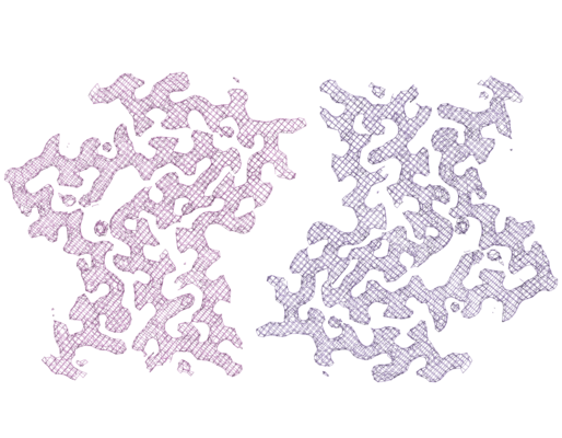 emd_20759.png emd_20759.png | 280.2 KB | ||
| Filedesc metadata | emd-20759.cif.gz | 5.6 KB | ||
| Archive directory |  http://ftp.pdbj.org/pub/emdb/structures/EMD-20759ftp://ftp.pdbj.org/pub/emdb/structures/EMD-20759 http://ftp.pdbj.org/pub/emdb/structures/EMD-20759ftp://ftp.pdbj.org/pub/emdb/structures/EMD-20759 | HTTPS FTP |
-Related structure data
| Related structure data |  6ufrMC M: atomic model generated by this map C: citing same article ( |
|---|---|
| Similar structure data | |
| EM raw data | EMPIAR-10494 (Title: The α-synuclein hereditary mutation E46K unlocks a more stable, pathogenic fibril structure Data size: 897.9 Data #1: Unaligned K3 frames from alpha-synuclein E46K amyloid fibrils [micrographs - multiframe]) |


-Links
| EMDB pages | EMDB (EBI/PDBe) / EMDataResource |
|---|---|
| Related items in Molecule of the Month |

-Map
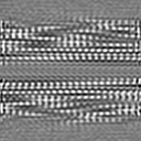
| File | Download / File: emd_20759.map.gz / Format: CCP4 / Size: 8 MB / Type: IMAGE STORED AS FLOATING POINT NUMBER (4 BYTES) | ||||||||||||||||||||||||||||||||||||||||||||||||||||||||||||||||||||
|---|---|---|---|---|---|---|---|---|---|---|---|---|---|---|---|---|---|---|---|---|---|---|---|---|---|---|---|---|---|---|---|---|---|---|---|---|---|---|---|---|---|---|---|---|---|---|---|---|---|---|---|---|---|---|---|---|---|---|---|---|---|---|---|---|---|---|---|---|---|
| Projections & slices | Image control
Images are generated by Spider. | ||||||||||||||||||||||||||||||||||||||||||||||||||||||||||||||||||||
| Voxel size | X=Y=Z: 0.838 Å | ||||||||||||||||||||||||||||||||||||||||||||||||||||||||||||||||||||
| Density |
| ||||||||||||||||||||||||||||||||||||||||||||||||||||||||||||||||||||
| Symmetry | Space group: 1 | ||||||||||||||||||||||||||||||||||||||||||||||||||||||||||||||||||||
| Details | EMDB XML:
CCP4 map header:
| ||||||||||||||||||||||||||||||||||||||||||||||||||||||||||||||||||||
 X (Sec.)
 Y (Row.)
 Z (Col.)


-Supplemental data
- Sample components
-Entire : Recombinantly assembled E46K alpha-synuclein fibril
| Entire | Name: Recombinantly assembled E46K alpha-synuclein fibril |
|---|---|
| Components |
|
-Supramolecule #1: Recombinantly assembled E46K alpha-synuclein fibril
| Supramolecule | Name: Recombinantly assembled E46K alpha-synuclein fibril / type: organelle_or_cellular_component / ID: 1 / Parent: 0 / Macromolecule list: #1 |
|---|---|
| Source (natural) | Organism: Homo sapiens (human) |
-Macromolecule #1: Alpha-synuclein
| Macromolecule | Name: Alpha-synuclein / type: protein_or_peptide / ID: 1 / Number of copies: 10 / Enantiomer: LEVO |
|---|---|
| Source (natural) | Organism: Homo sapiens (human) |
| Molecular weight | Theoretical: 14.476175 KDa |
| Recombinant expression | Organism:  |
| Sequence | String: MDVFMKGLSK AKEGVVAAAE KTKQGVAEAA GKTKEGVLYV GSKTKKGVVH GVATVAEKTK EQVTNVGGAV VTGVTAVAQK TVEGAGSIA AATGFVKKDQ LGKNEEGAPQ EGILEDMPVD PDNEAYEMPS EEGYQDYEPE A UniProtKB: Alpha-synuclein |
-Macromolecule #2: water
| Macromolecule | Name: water / type: ligand / ID: 2 / Number of copies: 90 / Formula: HOH |
|---|---|
| Molecular weight | Theoretical: 18.015 Da |
| Chemical component information |  ChemComp-HOH: |
-Experimental details
-Structure determination
| Method | cryo EM |
|---|---|
 Processing Processing | helical reconstruction |
| Aggregation state | filament |
-Sample preparation
| Buffer | pH: 7 |
|---|---|
| Vitrification | Cryogen name: ETHANE |
- Electron microscopy
| Microscope | FEI TITAN KRIOS |
|---|---|
| Image recording | Film or detector model: GATAN K2 SUMMIT (4k x 4k) / Detector mode: SUPER-RESOLUTION / Average electron dose: 36.0 e/Å2 |
| Electron beam | Acceleration voltage: 300 kV / Electron source:  FIELD EMISSION GUN FIELD EMISSION GUN |
| Electron optics | Illumination mode: FLOOD BEAM / Imaging mode: BRIGHT FIELD |
| Experimental equipment |  Model: Titan Krios / Image courtesy: FEI Company |

