 Movie
Movie Controller

[English] 日本語
 Yorodumi
- EMDB-20522: Cryo-EM structure of full-length insulin receptor bound to 4 insu... -
+ Open data

- Basic information
| Entry | Database: EMDB / ID: EMD-20522 | |||||||||
|---|---|---|---|---|---|---|---|---|---|---|
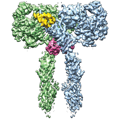
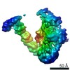
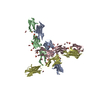
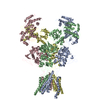
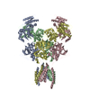
| Title | Cryo-EM structure of full-length insulin receptor bound to 4 insulin. 3D refinement was focused on the extracellular region. | |||||||||
 Map data Map data | Cryo-EM structure of full-length insulin receptor bound to 4 insulin. 3D refinement was focused on the entire extracellular region. | |||||||||
 Sample Sample |
| |||||||||
 Keywords Keywords | insulin receptor / insulin / SIGNALING PROTEIN-HORMONE complex | |||||||||
| Function / homology |  Function and homology information Function and homology informationcellular response to oxygen-containing compound / regulation of female gonad development / positive regulation of meiotic cell cycle / insulin-like growth factor II binding / positive regulation of developmental growth / male sex determination / insulin receptor complex / insulin-like growth factor I binding / insulin receptor activity / positive regulation of protein-containing complex disassembly ...cellular response to oxygen-containing compound / regulation of female gonad development / positive regulation of meiotic cell cycle / insulin-like growth factor II binding / positive regulation of developmental growth / male sex determination / insulin receptor complex / insulin-like growth factor I binding / insulin receptor activity / positive regulation of protein-containing complex disassembly / exocrine pancreas development / dendritic spine maintenance / adrenal gland development / insulin binding / cargo receptor activity / PTB domain binding / Signaling by Insulin receptor / IRS activation / neuronal cell body membrane / positive regulation of respiratory burst / amyloid-beta clearance / regulation of embryonic development / insulin receptor substrate binding / positive regulation of receptor internalization / epidermis development / positive regulation of glycogen biosynthetic process / Signal attenuation / protein kinase activator activity / transport across blood-brain barrier / phosphatidylinositol 3-kinase binding / heart morphogenesis / Insulin receptor recycling / insulin-like growth factor receptor binding / neuron projection maintenance / positive regulation of mitotic nuclear division / Insulin receptor signalling cascade / receptor-mediated endocytosis / dendrite membrane / positive regulation of glycolytic process / positive regulation of D-glucose import across plasma membrane / learning / hormone activity / receptor protein-tyrosine kinase / caveola / receptor internalization / cellular response to growth factor stimulus / male gonad development / glucose metabolic process / cellular response to insulin stimulus / memory / positive regulation of nitric oxide biosynthetic process / insulin receptor signaling pathway / protein autophosphorylation / late endosome / glucose homeostasis / amyloid-beta binding / PI5P, PP2A and IER3 Regulate PI3K/AKT Signaling / protein tyrosine kinase activity / positive regulation of MAPK cascade / positive regulation of canonical NF-kappaB signal transduction / positive regulation of phosphatidylinositol 3-kinase/protein kinase B signal transduction / lysosome / signaling receptor complex / endosome membrane / positive regulation of cell migration / G protein-coupled receptor signaling pathway / external side of plasma membrane / protein domain specific binding / axon / positive regulation of cell population proliferation / symbiont entry into host cell / regulation of DNA-templated transcription / positive regulation of DNA-templated transcription / GTP binding / protein-containing complex binding / extracellular exosome / extracellular region / ATP binding / membrane / identical protein binding / plasma membrane Similarity search - Function | |||||||||
| Biological species |  Homo sapiens (human) Homo sapiens (human) | |||||||||
| Method | single particle reconstruction / cryo EM / Resolution: 3.2 Å | |||||||||
 Authors Authors | Uchikawa E / Choi E | |||||||||
 Citation Citation | Journal: Elife / Year: 2019 Title: Activation mechanism of the insulin receptor revealed by cryo-EM structure of the fully liganded receptor-ligand complex. Authors: Emiko Uchikawa / Eunhee Choi / Guijun Shang / Hongtao Yu / Xiao-Chen Bai /  Abstract: Insulin signaling controls metabolic homeostasis. Here, we report the cryo-EM structure of full-length insulin receptor (IR) and insulin complex in the active state. This structure unexpectedly ...Insulin signaling controls metabolic homeostasis. Here, we report the cryo-EM structure of full-length insulin receptor (IR) and insulin complex in the active state. This structure unexpectedly reveals that maximally four insulins can bind the 'T'-shaped IR dimer at four distinct sites related by 2-fold symmetry. Insulins 1 and 1' bind to sites 1 and 1', formed by L1 of one IR protomer and α-CT and FnIII-1 of the other. Insulins 2 and 2' bind to sites 2 and 2' on FnIII-1 of each protomer. Mutagenesis and cellular assays show that both sites 1 and 2 are required for optimal insulin binding and IR activation. We further identify a homotypic FnIII-2-FnIII-2 interaction in mediating the dimerization of membrane proximal domains in the active IR dimer. Our results indicate that binding of multiple insulins at two distinct types of sites disrupts the autoinhibited apo-IR dimer and stabilizes the active dimer. | |||||||||
| History |
|
- Structure visualization
| Movie |
Movie viewer |
|---|---|
| Structure viewer | EM map: SurfViewMolmilJmol/JSmol |
| Supplemental images |
- Downloads & links
-EMDB archive
| Map data | emd_20522.map.gz | 96.2 MB | EMDB map data format | |
|---|---|---|---|---|
| Header (meta data) | emd-20522-v30.xmlemd-20522.xml | 13.8 KB 13.8 KB | Display Display | EMDB header |
| Images |  emd_20522.png emd_20522.png | 179.7 KB | ||
| Filedesc metadata | emd-20522.cif.gz | 6.1 KB | ||
| Archive directory |  http://ftp.pdbj.org/pub/emdb/structures/EMD-20522ftp://ftp.pdbj.org/pub/emdb/structures/EMD-20522 http://ftp.pdbj.org/pub/emdb/structures/EMD-20522ftp://ftp.pdbj.org/pub/emdb/structures/EMD-20522 | HTTPS FTP |
-Related structure data
| Related structure data |  6pxvMC  6pxwC C: citing same article ( M: atomic model generated by this map |
|---|---|
| Similar structure data |


-Links
| EMDB pages | EMDB (EBI/PDBe) / EMDataResource |
|---|---|
| Related items in Molecule of the Month |


-Map
| File | Download / File: emd_20522.map.gz / Format: CCP4 / Size: 103 MB / Type: IMAGE STORED AS FLOATING POINT NUMBER (4 BYTES) | ||||||||||||||||||||||||||||||||||||||||||||||||||||||||||||||||||||
|---|---|---|---|---|---|---|---|---|---|---|---|---|---|---|---|---|---|---|---|---|---|---|---|---|---|---|---|---|---|---|---|---|---|---|---|---|---|---|---|---|---|---|---|---|---|---|---|---|---|---|---|---|---|---|---|---|---|---|---|---|---|---|---|---|---|---|---|---|---|
| Annotation | Cryo-EM structure of full-length insulin receptor bound to 4 insulin. 3D refinement was focused on the entire extracellular region. | ||||||||||||||||||||||||||||||||||||||||||||||||||||||||||||||||||||
| Projections & slices | Image control
Images are generated by Spider. | ||||||||||||||||||||||||||||||||||||||||||||||||||||||||||||||||||||
| Voxel size | X=Y=Z: 1.07 Å | ||||||||||||||||||||||||||||||||||||||||||||||||||||||||||||||||||||
| Density |
| ||||||||||||||||||||||||||||||||||||||||||||||||||||||||||||||||||||
| Symmetry | Space group: 1 | ||||||||||||||||||||||||||||||||||||||||||||||||||||||||||||||||||||
| Details | EMDB XML:
CCP4 map header:
| ||||||||||||||||||||||||||||||||||||||||||||||||||||||||||||||||||||
 Z (Sec.)
 Y (Row.)
 X (Col.)


-Supplemental data
- Sample components
-Entire : Full-length insulin receptor/insulin complex
| Entire | Name: Full-length insulin receptor/insulin complex |
|---|---|
| Components |
|
-Supramolecule #1: Full-length insulin receptor/insulin complex
| Supramolecule | Name: Full-length insulin receptor/insulin complex / type: complex / ID: 1 / Parent: 0 / Macromolecule list: all |
|---|---|
| Source (natural) | Organism: Homo sapiens (human) |
| Molecular weight | Theoretical: 336 KDa |
-Macromolecule #1: Insulin receptor
| Macromolecule | Name: Insulin receptor / type: protein_or_peptide / ID: 1 / Number of copies: 2 / Enantiomer: LEVO / EC number: receptor protein-tyrosine kinase |
|---|---|
| Source (natural) | Organism: Homo sapiens (human) |
| Molecular weight | Theoretical: 153.913312 KDa |
| Recombinant expression | Organism: Homo sapiens (human) |
| Sequence | String: HLYPGEVCPG MDIRNNLTRL HELENCSVIE GHLQILLMFK TRPEDFRDLS FPKLIMITDY LLLFRVYGLE SLKDLFPNLT VIRGSRLFF NYALVIFEMV HLKELGLYNL MNITRGSVRI EKNNELCYLA TIDWSRILDS VEDNYIVLNK DDNEECGDIC P GTAKGKTN ...String: HLYPGEVCPG MDIRNNLTRL HELENCSVIE GHLQILLMFK TRPEDFRDLS FPKLIMITDY LLLFRVYGLE SLKDLFPNLT VIRGSRLFF NYALVIFEMV HLKELGLYNL MNITRGSVRI EKNNELCYLA TIDWSRILDS VEDNYIVLNK DDNEECGDIC P GTAKGKTN CPATVINGQF VERCWTHSHC QKVCPTICKS HGCTAEGLCC HSECLGNCSQ PDDPTKCVAC RNFYLDGRCV ET CPPPYYH FQDWRCVNFS FCQDLHHKCK NSRRQGCHQY VIHNNKCIPE CPSGYTMNSS NLLCTPCLGP CPKVCHLLEG EKT IDSVTS AQELRGCTVI NGSLIINIRG GNNLAAELEA NLGLIEEISG YLKIRRSYAL VSLSFFRKLR LIRGETLEIG NYSF YALDN QNLRQLWDWS KHNLTITQGK LFFHYNPKLC LSEIHKMEEV SGTKGRQERN DIALKTNGDQ ASCENELLKF SYIRT SFDK ILLRWEPYWP PDFRDLLGFM LFYKEAPYQN VTEFDGQDAC GSNSWTVVDI DPPLRSNDPK SQNHPGWLMR GLKPWT QYA IFVKTLVTFS DERRTYGAKS DIIYVQTDAT NPSVPLDPIS VSNSSSQIIL KWKPPSDPNG NITHYLVFWE RQAEDSE LF ELDYCLKGLK LPSRTWSPPF ESEDSQKHNQ SEYEDSAGEC CSCPKTDSQI LKELEESSFR KTFEDYLHNV VFVPRPSR K RRSLGDVGNV TVAVPTVAAF PNTSSTSVPT SPEEHRPFEK VVNKESLVIS GLRHFTGYRI ELQACNQDTP EERCSVAAY VSARTMPEAK ADDIVGPVTH EIFENNVVHL MWQEPKEPNG LIVLYEVSYR RYGDEELHLC VSRKHFALER GCRLRGLSPG NYSVRIRAT SLAGNGSWTE PTYFYVTDYL DVPSNIAKII IGPLIFVFLF SVVIGSIYLF LRKRQPDGPL GPLYASSNPE F LTASDVFP CSVYVPDEWE VSREKITLLR ELGQGSFGMV YEGNARDIIK GEAETRVAVK TVNESASLRE RIEFLNEASV MK GFTCHHV VRLLGVVSKG QPTLVVMELM AHGDLKSYLR SLRPEAENNP GRPPPTLQEM IQMAAEIADG MAYLNAKKFV HRN LAARNC MVAHDFTVKI GDFGMTRDIY ETDYYRKGGK GLLPVRWMAP ESLKDGVFTT SSDMWSFGVV LWEITSLAEQ PYQG LSNEQ VLKFVMDGGY LDQPDNCPER VTDLMRMCWQ FNPKMRPTFL EIVNLLKDDL HPSFPEVSFF HSEENKAPES EELEM EFED MENVPLDRSS HCQREEAGGR DGGSSLGFKR SYEEHIPYTH MNGGKKNGAA ATAPRSNPSL ESSGLEVLFQ UniProtKB: Insulin receptor |
-Macromolecule #2: Insulin
| Macromolecule | Name: Insulin / type: protein_or_peptide / ID: 2 / Number of copies: 4 / Enantiomer: LEVO |
|---|---|
| Source (natural) | Organism: Homo sapiens (human) |
| Molecular weight | Theoretical: 8.380529 KDa |
| Recombinant expression | Organism: Homo sapiens (human) |
| Sequence | String: FVNQHLCGSH LVEALYLVCG ERGFFYTPKT RREAEDLQGS LQPLALEGSL QKRGIVEQCC TSICSLYQLE NYCN UniProtKB: Insulin |
-Experimental details
-Structure determination
| Method | cryo EM |
|---|---|
 Processing Processing | single particle reconstruction |
| Aggregation state | particle |
-Sample preparation
| Concentration | 7 mg/mL |
|---|---|
| Buffer | pH: 7.5 |
| Grid | Model: Quantifoil R1.2/1.3 / Material: GOLD / Mesh: 300 / Pretreatment - Type: GLOW DISCHARGE |
| Vitrification | Cryogen name: ETHANE / Chamber humidity: 100 % / Instrument: FEI VITROBOT MARK IV |
- Electron microscopy
| Microscope | FEI TITAN KRIOS |
|---|---|
| Image recording | Film or detector model: GATAN K2 IS (4k x 4k) / Average exposure time: 15.0 sec. / Average electron dose: 50.0 e/Å2 |
| Electron beam | Acceleration voltage: 300 kV / Electron source:  FIELD EMISSION GUN FIELD EMISSION GUN |
| Electron optics | C2 aperture diameter: 70.0 µm / Illumination mode: FLOOD BEAM / Imaging mode: BRIGHT FIELD / Cs: 2.7 mm |
| Sample stage | Specimen holder model: FEI TITAN KRIOS AUTOGRID HOLDER / Cooling holder cryogen: NITROGEN |
| Experimental equipment |  Model: Titan Krios / Image courtesy: FEI Company |
+Image processing
-Atomic model buiding 1
| Refinement | Space: REAL / Protocol: FLEXIBLE FIT |
|---|---|
| Output model | PDB-6pxv: |
