Movie
Movie Controller


+ Open data

- Basic information
| Entry | Database: EMDB / ID: EMD-20268 | |||||||||
|---|---|---|---|---|---|---|---|---|---|---|
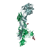
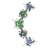
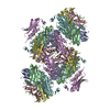
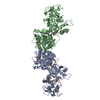
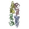
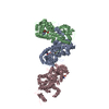
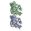
| Title | Chikungunya virus p62/E1 complex | |||||||||
 Map data Map data | Chikungunya virus p62/E1 complex | |||||||||
 Sample Sample |
| |||||||||
| Biological species |   Chikungunya virus Chikungunya virus | |||||||||
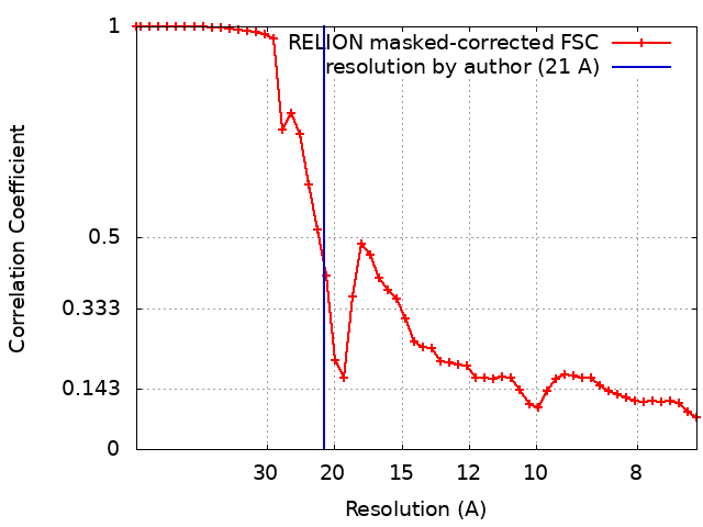
| Method | single particle reconstruction / negative staining / Resolution: 21.0 Å | |||||||||
 Authors Authors | Pallesen J / Ward AB | |||||||||
 Citation Citation | Journal: PLoS Pathog / Year: 2019 Title: Human monoclonal antibodies against chikungunya virus target multiple distinct epitopes in the E1 and E2 glycoproteins. Authors: Jose A Quiroz / Ryan J Malonis / Larissa B Thackray / Courtney A Cohen / Jesper Pallesen / Rohit K Jangra / Rebecca S Brown / Daniel Hofmann / Frederick W Holtsberg / Sergey Shulenin / ...Authors: Jose A Quiroz / Ryan J Malonis / Larissa B Thackray / Courtney A Cohen / Jesper Pallesen / Rohit K Jangra / Rebecca S Brown / Daniel Hofmann / Frederick W Holtsberg / Sergey Shulenin / Elisabeth K Nyakatura / Lorellin A Durnell / Vinayak Rayannavar / Johanna P Daily / Andrew B Ward / M Javad Aman / John M Dye / Kartik Chandran / Michael S Diamond / Margaret Kielian / Jonathan R Lai /  Abstract: Chikungunya virus (CHIKV) is a mosquito-transmitted alphavirus that causes persistent arthritis in a subset of human patients. We report the isolation and functional characterization of monoclonal ...Chikungunya virus (CHIKV) is a mosquito-transmitted alphavirus that causes persistent arthritis in a subset of human patients. We report the isolation and functional characterization of monoclonal antibodies (mAbs) from two patients infected with CHIKV in the Dominican Republic. Single B cell sorting yielded a panel of 46 human mAbs of diverse germline lineages that targeted epitopes within the E1 or E2 glycoproteins. MAbs that recognized either E1 or E2 proteins exhibited neutralizing activity. Viral escape mutations localized the binding epitopes for two E1 mAbs to sites within domain I or the linker between domains I and III; and for two E2 mAbs between the β-connector region and the B-domain. Two of the E2-specific mAbs conferred protection in vivo in a stringent lethal challenge mouse model of CHIKV infection, whereas the E1 mAbs did not. These results provide insight into human antibody response to CHIKV and identify candidate mAbs for therapeutic intervention. | |||||||||
| History |
|
- Structure visualization
| Movie |
 Movie viewer Movie viewer |
|---|---|
| Structure viewer | EM map: SurfViewMolmilJmol/JSmol |
| Supplemental images |

- Downloads & links
-EMDB archive
| Map data | emd_20268.map.gz | 6 MB | EMDB map data format | |
|---|---|---|---|---|
| Header (meta data) | emd-20268-v30.xmlemd-20268.xml | 14 KB 14 KB | Display Display | EMDB header |
| FSC (resolution estimation) | emd_20268_fsc.xml | 4.7 KB | Display | FSC data file |
| Images |  emd_20268.png emd_20268.png | 41 KB | ||
| Others | emd_20268_half_map_1.map.gzemd_20268_half_map_2.map.gz | 6 MB 6 MB | ||
| Archive directory |  http://ftp.pdbj.org/pub/emdb/structures/EMD-20268ftp://ftp.pdbj.org/pub/emdb/structures/EMD-20268 http://ftp.pdbj.org/pub/emdb/structures/EMD-20268ftp://ftp.pdbj.org/pub/emdb/structures/EMD-20268 | HTTPS FTP |
-Related structure data
| Related structure data | C: citing same article ( |
|---|---|
| Similar structure data |


-Links
| EMDB pages | EMDB (EBI/PDBe) / EMDataResource |
|---|
-Map
| File | Download / File: emd_20268.map.gz / Format: CCP4 / Size: 8 MB / Type: IMAGE STORED AS FLOATING POINT NUMBER (4 BYTES) | ||||||||||||||||||||||||||||||||||||||||||||||||||||||||||||||||||||
|---|---|---|---|---|---|---|---|---|---|---|---|---|---|---|---|---|---|---|---|---|---|---|---|---|---|---|---|---|---|---|---|---|---|---|---|---|---|---|---|---|---|---|---|---|---|---|---|---|---|---|---|---|---|---|---|---|---|---|---|---|---|---|---|---|---|---|---|---|---|
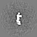
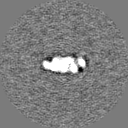
| Annotation | Chikungunya virus p62/E1 complex | ||||||||||||||||||||||||||||||||||||||||||||||||||||||||||||||||||||
| Projections & slices | Image control
Images are generated by Spider. | ||||||||||||||||||||||||||||||||||||||||||||||||||||||||||||||||||||
| Voxel size | X=Y=Z: 3.58 Å | ||||||||||||||||||||||||||||||||||||||||||||||||||||||||||||||||||||
| Density |
| ||||||||||||||||||||||||||||||||||||||||||||||||||||||||||||||||||||
| Symmetry | Space group: 1 | ||||||||||||||||||||||||||||||||||||||||||||||||||||||||||||||||||||
| Details | EMDB XML:
CCP4 map header:
| ||||||||||||||||||||||||||||||||||||||||||||||||||||||||||||||||||||
 Z (Sec.)
 Y (Row.)
 X (Col.)


-Supplemental data
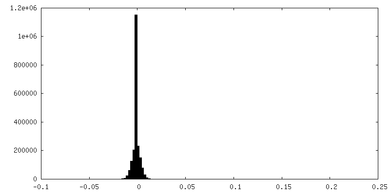
-Half map: Chikungunya virus p62/E1 complex, half map 1
| File | emd_20268_half_map_1.map | ||||||||||||
|---|---|---|---|---|---|---|---|---|---|---|---|---|---|
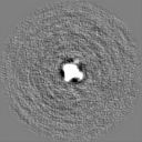
| Annotation | Chikungunya virus p62/E1 complex, half map 1 | ||||||||||||
| Projections & Slices |
| ||||||||||||
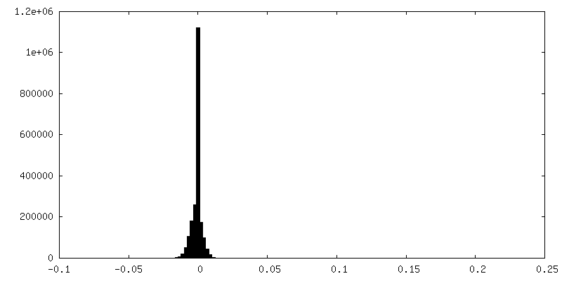
| Density Histograms |


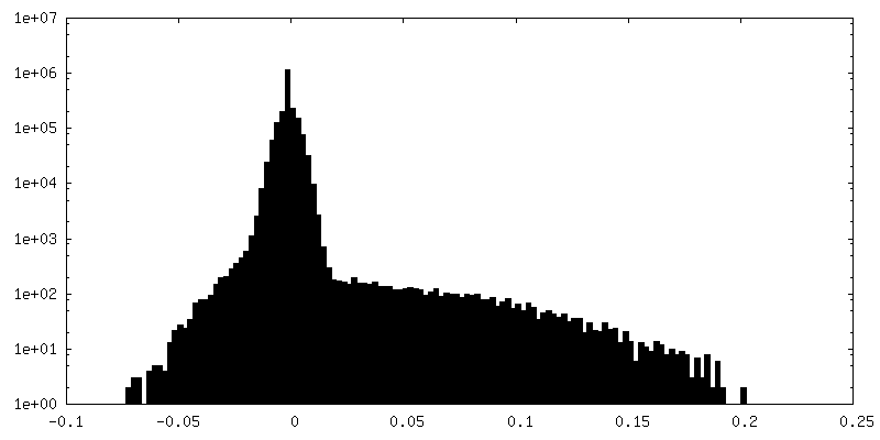
-Half map: Chikungunya virus p62/E1 complex, half map 2
| File | emd_20268_half_map_2.map | ||||||||||||
|---|---|---|---|---|---|---|---|---|---|---|---|---|---|
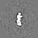
| Annotation | Chikungunya virus p62/E1 complex, half map 2 | ||||||||||||
| Projections & Slices |
| ||||||||||||
| Density Histograms |


- Sample components
-Entire : Chikungunya virus p62/E1 complex
| Entire | Name: Chikungunya virus p62/E1 complex |
|---|---|
| Components |
|
-Supramolecule #1: Chikungunya virus p62/E1 complex
| Supramolecule | Name: Chikungunya virus p62/E1 complex / type: complex / ID: 1 / Parent: 0 |
|---|---|
| Source (natural) | Organism: Chikungunya virus |
| Recombinant expression | Organism:  Homo sapiens (human) Homo sapiens (human) |
| Molecular weight | Theoretical: 85 KDa |
-Experimental details
-Structure determination
| Method | negative staining |
|---|---|
 Processing Processing | single particle reconstruction |
| Aggregation state | particle |
-Sample preparation
| Buffer | pH: 7 |
|---|---|
| Staining | Type: NEGATIVE / Material: uranyl formate |
| Grid | Details: unspecified |
- Electron microscopy
| Microscope | FEI TECNAI F20 |
|---|---|
| Image recording | Film or detector model: FEI EAGLE (4k x 4k) / Average electron dose: 30.0 e/Å2 |
| Electron beam | Acceleration voltage: 200 kV / Electron source:  FIELD EMISSION GUN FIELD EMISSION GUN |
| Electron optics | Illumination mode: FLOOD BEAM / Imaging mode: BRIGHT FIELD / Nominal defocus max: 1.5 µm / Nominal defocus min: 1.5 µm / Nominal magnification: 62000 |
| Experimental equipment |  Model: Tecnai F20 / Image courtesy: FEI Company |