 Movie
Movie Controller


+ Open data

- Basic information
| Entry | Database: EMDB / ID: EMD-20062 | |||||||||
|---|---|---|---|---|---|---|---|---|---|---|
| Title | Fractal-like assemblies of designed AtzA and AtzC proteins | |||||||||
 Map data Map data | Subtomogram of a small AtzA-AtzC assembly | |||||||||
 Sample Sample |
| |||||||||
| Biological species |  Pseudomonas sp. (bacteria) Pseudomonas sp. (bacteria) | |||||||||
| Method | electron tomography / cryo EM | |||||||||
 Authors Authors | Dai W / Chen M / Khare S | |||||||||
| Funding support |  United States, 1 items United States, 1 items
| |||||||||
 Citation Citation | Journal: Nat Chem / Year: 2019 Title: Stimulus-responsive self-assembly of protein-based fractals by computational design. Authors: Nancy E Hernández / William A Hansen / Denzel Zhu / Maria E Shea / Marium Khalid / Viacheslav Manichev / Matthew Putnins / Muyuan Chen / Anthony G Dodge / Lu Yang / Ileana Marrero-Berríos ...Authors: Nancy E Hernández / William A Hansen / Denzel Zhu / Maria E Shea / Marium Khalid / Viacheslav Manichev / Matthew Putnins / Muyuan Chen / Anthony G Dodge / Lu Yang / Ileana Marrero-Berríos / Melissa Banal / Phillip Rechani / Torgny Gustafsson / Leonard C Feldman / Sang-Hyuk Lee / Lawrence P Wackett / Wei Dai / Sagar D Khare / Abstract: Fractal topologies, which are statistically self-similar over multiple length scales, are pervasive in nature. The recurrence of patterns in fractal-shaped branched objects, such as trees, lungs and ...Fractal topologies, which are statistically self-similar over multiple length scales, are pervasive in nature. The recurrence of patterns in fractal-shaped branched objects, such as trees, lungs and sponges, results in a high surface area to volume ratio, which provides key functional advantages including molecular trapping and exchange. Mimicking these topologies in designed protein-based assemblies could provide access to functional biomaterials. Here we describe a computational design approach for the reversible self-assembly of proteins into tunable supramolecular fractal-like topologies in response to phosphorylation. Guided by atomic-resolution models, we develop fusions of Src homology 2 (SH2) domain or a phosphorylatable SH2-binding peptide, respectively, to two symmetric, homo-oligomeric proteins. Mixing the two designed components resulted in a variety of dendritic, hyperbranched and sponge-like topologies that are phosphorylation-dependent and self-similar over three decades (~10 nm-10 μm) of length scale, in agreement with models from multiscale computational simulations. Designed assemblies perform efficient phosphorylation-dependent capture and release of cargo proteins. | |||||||||
| History |
|
- Structure visualization
| Movie |
 Movie viewer Movie viewer |
|---|---|
| Structure viewer | EM map: SurfViewMolmilJmol/JSmol |
| Supplemental images |

- Downloads & links
-EMDB archive
| Map data | emd_20062.map.gz | 941 KB | EMDB map data format | |
|---|---|---|---|---|
| Header (meta data) | emd-20062-v30.xmlemd-20062.xml | 11.1 KB 11.1 KB | Display Display | EMDB header |
| Images | 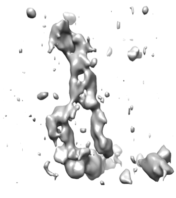 emd_20062.png emd_20062.png | 81 KB | ||
| Archive directory |  http://ftp.pdbj.org/pub/emdb/structures/EMD-20062ftp://ftp.pdbj.org/pub/emdb/structures/EMD-20062 http://ftp.pdbj.org/pub/emdb/structures/EMD-20062ftp://ftp.pdbj.org/pub/emdb/structures/EMD-20062 | HTTPS FTP |
-Related structure data

-Links
| EMDB pages | EMDB (EBI/PDBe) / EMDataResource |
|---|---|
| Related items in Molecule of the Month |

-Map
| File | Download / File: emd_20062.map.gz / Format: CCP4 / Size: 1012.7 KB / Type: IMAGE STORED AS FLOATING POINT NUMBER (4 BYTES) | ||||||||||||||||||||||||||||||||||||||||||||||||||||||||||||||||||||
|---|---|---|---|---|---|---|---|---|---|---|---|---|---|---|---|---|---|---|---|---|---|---|---|---|---|---|---|---|---|---|---|---|---|---|---|---|---|---|---|---|---|---|---|---|---|---|---|---|---|---|---|---|---|---|---|---|---|---|---|---|---|---|---|---|---|---|---|---|---|
| Annotation | Subtomogram of a small AtzA-AtzC assembly | ||||||||||||||||||||||||||||||||||||||||||||||||||||||||||||||||||||
| Projections & slices | Image control
Images are generated by Spider. generated in cubic-lattice coordinate | ||||||||||||||||||||||||||||||||||||||||||||||||||||||||||||||||||||
| Voxel size | X=Y=Z: 14 Å | ||||||||||||||||||||||||||||||||||||||||||||||||||||||||||||||||||||
| Density |
| ||||||||||||||||||||||||||||||||||||||||||||||||||||||||||||||||||||
| Symmetry | Space group: 1 | ||||||||||||||||||||||||||||||||||||||||||||||||||||||||||||||||||||
| Details | EMDB XML:
CCP4 map header:
| ||||||||||||||||||||||||||||||||||||||||||||||||||||||||||||||||||||
 Z (Sec.)
 Y (Row.)
 X (Col.)


-Supplemental data
- Sample components
-Entire : Designed proteins AtzA-pY:AtzC-SH2
| Entire | Name: Designed proteins AtzA-pY:AtzC-SH2 |
|---|---|
| Components |
|
-Supramolecule #1: Designed proteins AtzA-pY:AtzC-SH2
| Supramolecule | Name: Designed proteins AtzA-pY:AtzC-SH2 / type: complex / ID: 1 / Parent: 0 |
|---|---|
| Source (natural) | Organism: Pseudomonas sp. (bacteria) |
| Recombinant expression | Organism: |
-Experimental details
-Structure determination
| Method | cryo EM |
|---|---|
 Processing Processing | electron tomography |
| Aggregation state | particle |
-Sample preparation
| Buffer | pH: 7 |
|---|---|
| Grid | Model: Quantifoil R2/1 / Material: COPPER / Mesh: 200 / Support film - Material: CARBON / Support film - topology: HOLEY / Support film - Film thickness: 0.5 nm / Pretreatment - Type: GLOW DISCHARGE / Pretreatment - Atmosphere: AIR / Pretreatment - Pressure: 101.325 kPa |
| Vitrification | Cryogen name: ETHANE / Chamber humidity: 90 % / Chamber temperature: 293 K / Instrument: LEICA EM GP |
| Sectioning | Other: NO SECTIONING |
| Fiducial marker | Manufacturer: EMS / Diameter: 10 nm |
- Electron microscopy
| Microscope | FEI TECNAI ARCTICA |
|---|---|
| Temperature | Min: 93.0 K / Max: 110.0 K |
| Specialist optics | Energy filter - Name: GIF Quantum LS / Energy filter - Slit width: 20 eV |
| Image recording | Film or detector model: GATAN K2 QUANTUM (4k x 4k) / Detector mode: COUNTING / Average exposure time: 2.0 sec. / Average electron dose: 1.0 e/Å2 |
| Electron beam | Acceleration voltage: 200 kV / Electron source:  FIELD EMISSION GUN FIELD EMISSION GUN |
| Electron optics | C2 aperture diameter: 100.0 µm / Calibrated defocus max: 6.0 µm / Calibrated defocus min: 4.0 µm / Calibrated magnification: 39000 / Illumination mode: FLOOD BEAM / Imaging mode: BRIGHT FIELD / Cs: 2.7 mm / Nominal defocus max: 6.0 µm / Nominal defocus min: 4.0 µm / Nominal magnification: 39000 |
| Sample stage | Specimen holder model: FEI TITAN KRIOS AUTOGRID HOLDER / Cooling holder cryogen: NITROGEN |
| Experimental equipment |  Model: Talos Arctica / Image courtesy: FEI Company |
-Image processing
| Final reconstruction | Algorithm: BACK PROJECTION / Software - Name: EMAN2 / Number images used: 400 |
|---|
-Atomic model buiding 1
| Refinement | Protocol: AB INITIO MODEL |
|---|
