 ムービー
ムービー コントローラー


+ データを開く

- 基本情報
基本情報
| 登録情報 | データベース: EMDB / ID: EMD-2004 | |||||||||
|---|---|---|---|---|---|---|---|---|---|---|
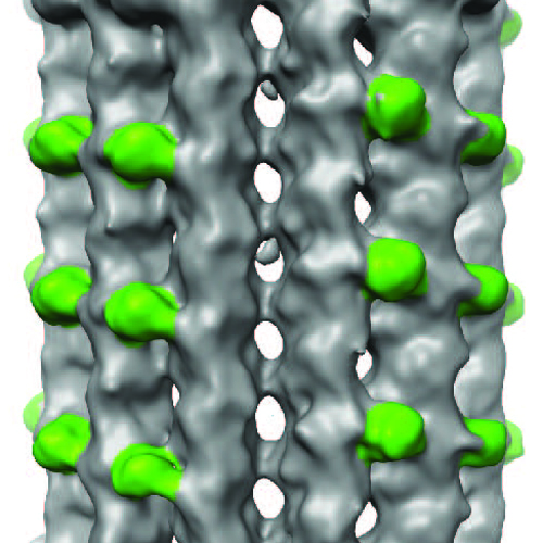
| タイトル | Asymmetric reconstruction of 13-protofilament GTPgammaS microtubules decorated with Mal3 CH domain | |||||||||
 マップデータ マップデータ | Asymmetric reconstruction of 13-protofilament GTPgammaS microtubules decorated with Mal3 CH domain | |||||||||
 試料 試料 |
| |||||||||
 キーワード キーワード | cytoskeleton / GTPase / end binding / calponin homology | |||||||||
| 生物種 |  | |||||||||
| 手法 | 単粒子再構成法 / クライオ電子顕微鏡法 / 解像度: 15.0 Å | |||||||||
 データ登録者 データ登録者 | Maurer SP / Fourniol FJ / Bohner G / Moores CA / Surrey T | |||||||||
 引用 引用 | ジャーナル: Cell / 年: 2012 タイトル: EBs recognize a nucleotide-dependent structural cap at growing microtubule ends. 著者: Sebastian P Maurer / Franck J Fourniol / Gergő Bohner / Carolyn A Moores / Thomas Surrey /  要旨: Growing microtubule ends serve as transient binding platforms for essential proteins that regulate microtubule dynamics and their interactions with cellular substructures. End-binding proteins (EBs) ...Growing microtubule ends serve as transient binding platforms for essential proteins that regulate microtubule dynamics and their interactions with cellular substructures. End-binding proteins (EBs) autonomously recognize an extended region at growing microtubule ends with unknown structural characteristics and then recruit other factors to the dynamic end structure. Using cryo-electron microscopy, subnanometer single-particle reconstruction, and fluorescence imaging, we present a pseudoatomic model of how the calponin homology (CH) domain of the fission yeast EB Mal3 binds to the end regions of growing microtubules. The Mal3 CH domain bridges protofilaments except at the microtubule seam. By binding close to the exchangeable GTP-binding site, the CH domain is ideally positioned to sense the microtubule's nucleotide state. The same microtubule-end region is also a stabilizing structural cap protecting the microtubule from depolymerization. This insight supports a common structural link between two important biological phenomena, microtubule dynamic instability and end tracking. | |||||||||
| 履歴 |
|
- 構造の表示
構造の表示
| ムービー |
 ムービービューア ムービービューア |
|---|---|
| 構造ビューア | EMマップ: SurfViewMolmilJmol/JSmol |
| 添付画像 |
- ダウンロードとリンク
-EMDBアーカイブ
| マップデータ | emd_2004.map.gz | 56.5 MB | EMDBマップデータ形式 | |
|---|---|---|---|---|
| ヘッダ (付随情報) | emd-2004-v30.xmlemd-2004.xml | 10.3 KB 10.3 KB | 表示 表示 | EMDBヘッダ |
| 画像 |  2004.jpg 2004.jpg | 846 KB | ||
| アーカイブディレクトリ |  http://ftp.pdbj.org/pub/emdb/structures/EMD-2004ftp://ftp.pdbj.org/pub/emdb/structures/EMD-2004 http://ftp.pdbj.org/pub/emdb/structures/EMD-2004ftp://ftp.pdbj.org/pub/emdb/structures/EMD-2004 | HTTPS FTP |
-関連構造データ

-リンク
| EMDBのページ | EMDB (EBI/PDBe) / EMDataResource |
|---|
-マップ
| ファイル | ダウンロード / ファイル: emd_2004.map.gz / 形式: CCP4 / 大きさ: 73.3 MB / タイプ: IMAGE STORED AS FLOATING POINT NUMBER (4 BYTES) | ||||||||||||||||||||||||||||||||||||||||||||||||||||||||||||||||||||
|---|---|---|---|---|---|---|---|---|---|---|---|---|---|---|---|---|---|---|---|---|---|---|---|---|---|---|---|---|---|---|---|---|---|---|---|---|---|---|---|---|---|---|---|---|---|---|---|---|---|---|---|---|---|---|---|---|---|---|---|---|---|---|---|---|---|---|---|---|---|
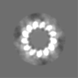
| 注釈 | Asymmetric reconstruction of 13-protofilament GTPgammaS microtubules decorated with Mal3 CH domain | ||||||||||||||||||||||||||||||||||||||||||||||||||||||||||||||||||||
| 投影像・断面図 | 画像のコントロール
画像は Spider により作成 | ||||||||||||||||||||||||||||||||||||||||||||||||||||||||||||||||||||
| ボクセルのサイズ | X=Y=Z: 2.2 Å | ||||||||||||||||||||||||||||||||||||||||||||||||||||||||||||||||||||
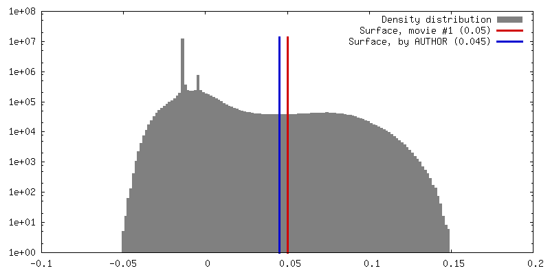
| 密度 |
| ||||||||||||||||||||||||||||||||||||||||||||||||||||||||||||||||||||
| 対称性 | 空間群: 1 | ||||||||||||||||||||||||||||||||||||||||||||||||||||||||||||||||||||
| 詳細 | EMDB XML:
CCP4マップ ヘッダ情報:
| ||||||||||||||||||||||||||||||||||||||||||||||||||||||||||||||||||||
 Z (Sec.)
 Y (Row.)
 X (Col.)


-添付データ
- 試料の構成要素
-全体 : 13-protofilament GTPgammaS microtubule decorated with monomeric Mal3
| 全体 | 名称: 13-protofilament GTPgammaS microtubule decorated with monomeric Mal3 |
|---|---|
| 要素 |
|
-超分子 #1000: 13-protofilament GTPgammaS microtubule decorated with monomeric Mal3
| 超分子 | 名称: 13-protofilament GTPgammaS microtubule decorated with monomeric Mal3 タイプ: sample / ID: 1000 詳細: tubulin was mixed with GMPCPP microtubule seeds, GTPgammaS and monomeric Mal3, and incubated 3-10min at 37degC 集合状態: 13-protofilament microtubule / Number unique components: 3 |
|---|
-分子 #1: Alpha-beta tubulin dimer
| 分子 | 名称: Alpha-beta tubulin dimer / タイプ: protein_or_peptide / ID: 1 / Name.synonym: Alpha-beta tubulin dimer / 集合状態: Dimer / 組換発現: No |
|---|---|
| 由来(天然) | 生物種: |
| 分子量 | 実験値: 100 KDa / 理論値: 100 KDa |
-分子 #3: Mal3
| 分子 | 名称: Mal3 / タイプ: protein_or_peptide / ID: 3 / Name.synonym: Mal3 / 集合状態: monomer / 組換発現: Yes |
|---|---|
| 由来(天然) | 生物種: Saccharomyces pombe / 別称: fission yeast / 細胞中の位置: cytoplasmic |
| 分子量 | 実験値: 16 KDa / 理論値: 16 KDa |
| 組換発現 | 生物種:  |
-分子 #2: guanosine 5'-O-(gamma-thio)triphosphate
| 分子 | 名称: guanosine 5'-O-(gamma-thio)triphosphate / タイプ: ligand / ID: 2 / Name.synonym: GTPgammaS / 組換発現: No |
|---|---|
| 由来(天然) | 生物種: synthetic construct (人工物) |
-実験情報
-構造解析
| 手法 | クライオ電子顕微鏡法 |
|---|---|
 解析 解析 | 単粒子再構成法 |
| 試料の集合状態 | particle |
-試料調製
| 緩衝液 | pH: 6.8 / 詳細: 40mM Pipes, 1mM MgCl2, 1mM EGTA |
|---|---|
| グリッド | 詳細: 300 mesh lacey carbon grid |
| 凍結 | 凍結剤: ETHANE / チャンバー内湿度: 90 % / 装置: OTHER / 詳細: Vitrification instrument: Vitrobot (FEI) / 手法: Chamber at 37 degrees C, blot 2s |
- 電子顕微鏡法
| 顕微鏡 | FEI TECNAI F20 |
|---|---|
| 温度 | 最低: 88 K / 最高: 98 K / 平均: 93 K |
| アライメント法 | Legacy - 非点収差: Objective lens astigmatism was corrected at 150,000 times magnification |
| 撮影 | カテゴリ: CCD フィルム・検出器のモデル: GATAN ULTRASCAN 4000 (4k x 4k) 実像数: 162 / 平均電子線量: 17 e/Å2 / 詳細: sampling size 2.2 A per pixel |
| 電子線 | 加速電圧: 200 kV / 電子線源:  FIELD EMISSION GUN FIELD EMISSION GUN |
| 電子光学系 | 照射モード: FLOOD BEAM / 撮影モード: BRIGHT FIELD / Cs: 2.0 mm / 最大 デフォーカス(公称値): 3.6 µm / 最小 デフォーカス(公称値): 0.7 µm / 倍率(公称値): 68000 |
| 試料ステージ | 試料ホルダー: Eucentric / 試料ホルダーモデル: OTHER |
| 実験機器 |  モデル: Tecnai F20 / 画像提供: FEI Company |
-画像解析
| CTF補正 | 詳細: FREALIGN |
|---|---|
| 最終 再構成 | 想定した対称性 - 点群: C1 (非対称) / アルゴリズム: OTHER / 解像度のタイプ: BY AUTHOR / 解像度: 15.0 Å / 解像度の算出法: FSC 0.5 CUT-OFF / ソフトウェア - 名称: SPIDER, FREALIGN 詳細: C1 map calculated from approximately 11000 one-dimer-long microtubule segments |
