National Health and Medical Research Council (Australia)
1146403
Australia
Citation
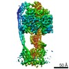
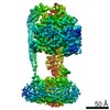
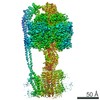
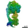
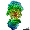
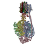
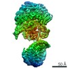
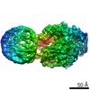
Journal: Elife / Year: 2019 Title: Cryo-EM reveals distinct conformations of ATP synthase on exposure to ATP. Authors: Meghna Sobti / Robert Ishmukhametov / James C Bouwer / Anita Ayer / Cacang Suarna / Nicola J Smith / Mary Christie / Roland Stocker / Thomas M Duncan / Alastair G Stewart / Abstract: ATP synthase produces the majority of cellular energy in most cells. We have previously reported cryo-EM maps of autoinhibited ATP synthase imaged without addition of nucleotide (Sobti et al. 2016), ...ATP synthase produces the majority of cellular energy in most cells. We have previously reported cryo-EM maps of autoinhibited ATP synthase imaged without addition of nucleotide (Sobti et al. 2016), indicating that the subunit ε engages the α, β and γ subunits to lock the enzyme and prevent functional rotation. Here we present multiple cryo-EM reconstructions of the enzyme frozen after the addition of MgATP to identify the changes that occur when this ε inhibition is removed. The maps generated show that, after exposure to MgATP, ATP synthase adopts a different conformation with a catalytic subunit changing conformation substantially and the ε C-terminal domain transitioning via an intermediate 'half-up' state to a condensed 'down' state. This work provides direct evidence for unique conformational states that occur in ATP synthase when ATP binding prevents the ε C-terminal domain from entering the inhibitory 'up' state.
History
Deposition
Mar 19, 2019
-
Header (metadata) release
Jul 3, 2019
-
Map release
Jul 3, 2019
-
Update
Jul 3, 2019
-
Current status
Jul 3, 2019
Processing site: RCSB / Status: Released
-
Structure visualization
Movie
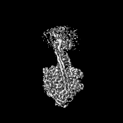
Surface view with section colored by density value
In the structure databanks used in Yorodumi, some data are registered as the other names, "COVID-19 virus" and "2019-nCoV". Here are the details of the virus and the list of structure data.
Jan 31, 2019. EMDB accession codes are about to change! (news from PDBe EMDB page)
EMDB accession codes are about to change! (news from PDBe EMDB page)
The allocation of 4 digits for EMDB accession codes will soon come to an end. Whilst these codes will remain in use, new EMDB accession codes will include an additional digit and will expand incrementally as the available range of codes is exhausted. The current 4-digit format prefixed with “EMD-” (i.e. EMD-XXXX) will advance to a 5-digit format (i.e. EMD-XXXXX), and so on. It is currently estimated that the 4-digit codes will be depleted around Spring 2019, at which point the 5-digit format will come into force.
The EM Navigator/Yorodumi systems omit the EMD- prefix.
Related info.:Q: What is EMD? / ID/Accession-code notation in Yorodumi/EM Navigator
Yorodumi is a browser for structure data from EMDB, PDB, SASBDB, etc.
This page is also the successor to EM Navigator detail page, and also detail information page/front-end page for Omokage search.
The word "yorodu" (or yorozu) is an old Japanese word meaning "ten thousand". "mi" (miru) is to see.
Related info.:EMDB / PDB / SASBDB / Comparison of 3 databanks / Yorodumi Search / Aug 31, 2016. New EM Navigator & Yorodumi / Yorodumi Papers / Jmol/JSmol / Function and homology information / Changes in new EM Navigator and Yorodumi
 Movie
Movie Controller


 Open data
 Basic information
 Map data
 Sample
 Authors
 Australia, 1 items
  Citation

 Structure visualization
 Movie viewer
 Downloads & links
 emd_20008.png
 http://ftp.pdbj.org/pub/emdb/structures/EMD-20008
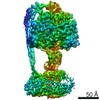


 Z (Sec.)
 Y (Row.)
 X (Col.)


 Sample components
 Processing
 Electron microscopy
 FIELD EMISSION GUN
