Journal: Nat Plants / Year: 2024 Title: Molecular basis of plastoquinone reduction in plant cytochrome bf. Authors: Sebastian Pintscher / Rafał Pietras / Bohun Mielecki / Mateusz Szwalec / Anna Wójcik-Augustyn / Paulina Indyka / Michał Rawski / Łukasz Koziej / Marcin Jaciuk / Grzegorz Ważny / ...Authors: Sebastian Pintscher / Rafał Pietras / Bohun Mielecki / Mateusz Szwalec / Anna Wójcik-Augustyn / Paulina Indyka / Michał Rawski / Łukasz Koziej / Marcin Jaciuk / Grzegorz Ważny / Sebastian Glatt / Artur Osyczka / Abstract: A multi-subunit enzyme, cytochrome bf (cytbf), provides the crucial link between photosystems I and II in the photosynthetic membranes of higher plants, transferring electrons between plastoquinone ...A multi-subunit enzyme, cytochrome bf (cytbf), provides the crucial link between photosystems I and II in the photosynthetic membranes of higher plants, transferring electrons between plastoquinone (PQ) and plastocyanin. The atomic structure of cytbf is known, but its detailed catalytic mechanism remains elusive. Here we present cryogenic electron microscopy structures of spinach cytbf at 1.9 Å and 2.2 Å resolution, revealing an unexpected orientation of the substrate PQ in the haem ligand niche that forms the PQ reduction site (Q). PQ, unlike Q inhibitors, is not in direct contact with the haem. Instead, a water molecule is coordinated by one of the carbonyl groups of PQ and can act as the immediate proton donor for PQ. In addition, we identify water channels that connect Q with the aqueous exterior of the enzyme, suggesting that the binding of PQ in Q displaces water through these channels. The structures confirm large movements of the head domain of the iron-sulfur protein (ISP-HD) towards and away from the plastoquinol oxidation site (Q) and define the unique position of ISP-HD when a Q inhibitor (2,5-dibromo-3-methyl-6-isopropylbenzoquinone) is bound. This work identifies key conformational states of cytbf, highlights fundamental differences between substrates and inhibitors and proposes a quinone-water exchange mechanism.
Supramolecule #1: cytochrome b6f complex with decylplastoquinol bound at quinone re...
Supramolecule
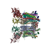
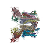
Name: cytochrome b6f complex with decylplastoquinol bound at quinone reduction site type: complex / ID: 1 / Parent: 0 / Macromolecule list: #1-#9 Details: cytochrome b6f complex incubated with reduced decyplastoquinone was mixed with plastocyanin and vitrified during turnover
In the structure databanks used in Yorodumi, some data are registered as the other names, "COVID-19 virus" and "2019-nCoV". Here are the details of the virus and the list of structure data.
Jan 31, 2019. EMDB accession codes are about to change! (news from PDBe EMDB page)
EMDB accession codes are about to change! (news from PDBe EMDB page)
The allocation of 4 digits for EMDB accession codes will soon come to an end. Whilst these codes will remain in use, new EMDB accession codes will include an additional digit and will expand incrementally as the available range of codes is exhausted. The current 4-digit format prefixed with “EMD-” (i.e. EMD-XXXX) will advance to a 5-digit format (i.e. EMD-XXXXX), and so on. It is currently estimated that the 4-digit codes will be depleted around Spring 2019, at which point the 5-digit format will come into force.
The EM Navigator/Yorodumi systems omit the EMD- prefix.
Related info.:Q: What is EMD? / ID/Accession-code notation in Yorodumi/EM Navigator
Yorodumi is a browser for structure data from EMDB, PDB, SASBDB, etc.
This page is also the successor to EM Navigator detail page, and also detail information page/front-end page for Omokage search.
The word "yorodu" (or yorozu) is an old Japanese word meaning "ten thousand". "mi" (miru) is to see.
Related info.:EMDB / PDB / SASBDB / Comparison of 3 databanks / Yorodumi Search / Aug 31, 2016. New EM Navigator & Yorodumi / Yorodumi Papers / Jmol/JSmol / Function and homology information / Changes in new EM Navigator and Yorodumi
 Movie
Movie Controller

 Yorodumi
 Open data
 Basic information
 Map data
 Sample
 Keywords
 Function and homology information
 Spinacia oleracea (spinach)
 Authors
 Poland, 3 items
  Citation
 Structure visualization
 Downloads & links
 emd_19939.png
 http://ftp.pdbj.org/pub/emdb/structures/EMD-19939
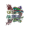


 Z (Sec.)
 Y (Row.)
 X (Col.)


 Sample components


 Processing
 Electron microscopy
 FIELD EMISSION GUN


