[日本語] English
 万見
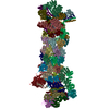
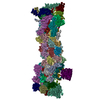
万見- EMDB-1992: The Saccharomyces cerevisiae 26S proteasome at subnanometer resolution -
+ データを開く
- 基本情報
基本情報
| 登録情報 | データベース: EMDB / ID: EMD-1992 | |||||||||
|---|---|---|---|---|---|---|---|---|---|---|
| タイトル | The Saccharomyces cerevisiae 26S proteasome at subnanometer resolution | |||||||||
 マップデータ マップデータ | 26 proteasome | |||||||||
 試料 試料 |
| |||||||||
 キーワード キーワード | 26S / 19S / proteasome / regulatory particle / ubiquitin recognition / deubiquitination / AAA-ATPase | |||||||||
| 機能・相同性 | proteasome regulatory particle / Proteasome, subunit alpha/beta 機能・相同性情報 機能・相同性情報 | |||||||||
| 生物種 |  | |||||||||
| 手法 | 単粒子再構成法 / クライオ電子顕微鏡法 / ネガティブ染色法 / 解像度: 9.0 Å | |||||||||
 データ登録者 データ登録者 | Lander GC / Estrin E / Matyskiela M / Bashore C / Nogales E / Martin A | |||||||||
 引用 引用 |  ジャーナル: Nature / 年: 2012 ジャーナル: Nature / 年: 2012タイトル: Complete subunit architecture of the proteasome regulatory particle. 著者: Gabriel C Lander / Eric Estrin / Mary E Matyskiela / Charlene Bashore / Eva Nogales / Andreas Martin /  要旨: The proteasome is the major ATP-dependent protease in eukaryotic cells, but limited structural information restricts a mechanistic understanding of its activities. The proteasome regulatory particle, ...The proteasome is the major ATP-dependent protease in eukaryotic cells, but limited structural information restricts a mechanistic understanding of its activities. The proteasome regulatory particle, consisting of the lid and base subcomplexes, recognizes and processes polyubiquitinated substrates. Here we used electron microscopy and a new heterologous expression system for the lid to delineate the complete subunit architecture of the regulatory particle from yeast. Our studies reveal the spatial arrangement of ubiquitin receptors, deubiquitinating enzymes and the protein unfolding machinery at subnanometre resolution, outlining the substrate's path to degradation. Unexpectedly, the ATPase subunits within the base unfoldase are arranged in a spiral staircase, providing insight into potential mechanisms for substrate translocation through the central pore. Large conformational rearrangements of the lid upon holoenzyme formation suggest allosteric regulation of deubiquitination. We provide a structural basis for the ability of the proteasome to degrade a diverse set of substrates and thus regulate vital cellular processes. | |||||||||
| 履歴 |
|
- 構造の表示
構造の表示
| ムービー |
 ムービービューア ムービービューア |
|---|---|
| 構造ビューア | EMマップ:  SurfView SurfView Molmil Molmil Jmol/JSmol Jmol/JSmol |
| 添付画像 |
- ダウンロードとリンク
-EMDBアーカイブ
| マップデータ |  emd_1992.map.gz emd_1992.map.gz | 2.8 MB |  EMDBマップデータ形式 EMDBマップデータ形式 | |
|---|---|---|---|---|
| ヘッダ (付随情報) |  emd-1992-v30.xml emd-1992-v30.xml emd-1992.xml emd-1992.xml | 10 KB 10 KB | 表示 表示 |  EMDBヘッダ EMDBヘッダ |
| 画像 |  emd-1992.png emd-1992.png | 199.4 KB | ||
| アーカイブディレクトリ |  http://ftp.pdbj.org/pub/emdb/structures/EMD-1992 http://ftp.pdbj.org/pub/emdb/structures/EMD-1992 ftp://ftp.pdbj.org/pub/emdb/structures/EMD-1992 ftp://ftp.pdbj.org/pub/emdb/structures/EMD-1992 | HTTPS FTP |
-関連構造データ
- リンク
リンク
| EMDBのページ |  EMDB (EBI/PDBe) / EMDB (EBI/PDBe) /  EMDataResource EMDataResource |
|---|
- マップ
マップ
| ファイル |  ダウンロード / ファイル: emd_1992.map.gz / 形式: CCP4 / 大きさ: 62.5 MB / タイプ: IMAGE STORED AS FLOATING POINT NUMBER (4 BYTES) ダウンロード / ファイル: emd_1992.map.gz / 形式: CCP4 / 大きさ: 62.5 MB / タイプ: IMAGE STORED AS FLOATING POINT NUMBER (4 BYTES) | ||||||||||||||||||||||||||||||||||||||||||||||||||||||||||||||||||||
|---|---|---|---|---|---|---|---|---|---|---|---|---|---|---|---|---|---|---|---|---|---|---|---|---|---|---|---|---|---|---|---|---|---|---|---|---|---|---|---|---|---|---|---|---|---|---|---|---|---|---|---|---|---|---|---|---|---|---|---|---|---|---|---|---|---|---|---|---|---|
| 注釈 | 26 proteasome | ||||||||||||||||||||||||||||||||||||||||||||||||||||||||||||||||||||
| 投影像・断面図 | 画像のコントロール
画像は Spider により作成 | ||||||||||||||||||||||||||||||||||||||||||||||||||||||||||||||||||||
| ボクセルのサイズ | X=Y=Z: 2.17 Å | ||||||||||||||||||||||||||||||||||||||||||||||||||||||||||||||||||||
| 密度 |
| ||||||||||||||||||||||||||||||||||||||||||||||||||||||||||||||||||||
| 対称性 | 空間群: 1 | ||||||||||||||||||||||||||||||||||||||||||||||||||||||||||||||||||||
| 詳細 | EMDB XML:
CCP4マップ ヘッダ情報:
| ||||||||||||||||||||||||||||||||||||||||||||||||||||||||||||||||||||
-添付データ
- 試料の構成要素
-全体 : 26S Proteasome
| 全体 | 名称: 26S Proteasome |
|---|---|
| 要素 |
|
-超分子 #1000: 26S Proteasome
| 超分子 | 名称: 26S Proteasome / タイプ: sample / ID: 1000 / 詳細: monodisperse / 集合状態: holoenzyme / Number unique components: 1 |
|---|---|
| 分子量 | 実験値: 1.5 MDa / 理論値: 1.5 MDa / 手法: Mass Spectrometry |
-分子 #1: 26S Proteasome
| 分子 | 名称: 26S Proteasome / タイプ: protein_or_peptide / ID: 1 / Name.synonym: 26S Proteasome / 詳細: Endogenous proteasome was purified from yeast / コピー数: 1 / 集合状態: monomer / 組換発現: No |
|---|---|
| 由来(天然) | 生物種:  |
| 分子量 | 実験値: 1.5 MDa / 理論値: 1.5 MDa |
| 配列 | GO: proteasome regulatory particle / InterPro: Proteasome, subunit alpha/beta |
-実験情報
-構造解析
| 手法 | ネガティブ染色法, クライオ電子顕微鏡法 |
|---|---|
 解析 解析 | 単粒子再構成法 |
| 試料の集合状態 | particle |
- 試料調製
試料調製
| 濃度 | 1.7 mg/mL |
|---|---|
| 緩衝液 | pH: 7.6 詳細: 20mM HEPES, 50mM NaCl, 50mM KCl, 1mM ATP, 1mM DTT, 0.05% NP40 |
| 染色 | タイプ: NEGATIVE 詳細: C-flat grids made hydrophilic with Solarus Plasma cleaner, 4 uL of sample applied, blotted in Vitrobot for 3 seconds with offset -1, plunged into liquid ethane |
| グリッド | 詳細: 400-mesh C-flats, 2um holes with 2um spacing (Protochips Inc.) |
| 凍結 | 凍結剤: ETHANE / チャンバー内湿度: 100 % / チャンバー内温度: 86 K / 装置: OTHER / 詳細: Vitrification instrument: Vitrobot / 手法: Blot 3 seconds with -2 offset |
- 電子顕微鏡法
| 顕微鏡 | FEI TECNAI 20 |
|---|---|
| 温度 | 最低: 78 K / 最高: 78 K / 平均: 78 K |
| アライメント法 | Legacy - 非点収差: objective lens astigmatism was corrected at 210,000 times magnification Legacy - Electron beam tilt params: 0 |
| 詳細 | Data acquired using Leginon |
| 日付 | 2011年9月11日 |
| 撮影 | カテゴリ: CCD フィルム・検出器のモデル: GENERIC GATAN (4k x 4k) 実像数: 9153 / 平均電子線量: 20 e/Å2 |
| 電子線 | 加速電圧: 120 kV / 電子線源:  FIELD EMISSION GUN FIELD EMISSION GUN |
| 電子光学系 | 照射モード: FLOOD BEAM / 撮影モード: BRIGHT FIELD / Cs: 2.2 mm / 最大 デフォーカス(公称値): 2.6 µm / 最小 デフォーカス(公称値): 1.2 µm / 倍率(公称値): 100000 |
| 試料ステージ | 試料ホルダー: Side-entry cryostage / 試料ホルダーモデル: GATAN LIQUID NITROGEN |
- 画像解析
画像解析
| 詳細 | Image processing performed in the Appion processing environment. 3D reconstruction performed using EMAN2 and SPARX libraries |
|---|---|
| CTF補正 | 詳細: whole micrograph |
| 最終 再構成 | 想定した対称性 - 点群: C2 (2回回転対称) / アルゴリズム: OTHER / 解像度のタイプ: BY AUTHOR / 解像度: 9.0 Å / 解像度の算出法: FSC 0.5 CUT-OFF / ソフトウェア - 名称: EMAN2 SPARX 詳細: Final map filtered to local resolution using the blocfilt function in Bsoft 使用した粒子像数: 93679 |
 ムービー
ムービー コントローラー


 Z (Sec.)
 Y (Row.)
 X (Col.)


