ムービー
ムービー コントローラー


+ データを開く

- 基本情報
基本情報
| 登録情報 |  | |||||||||
|---|---|---|---|---|---|---|---|---|---|---|
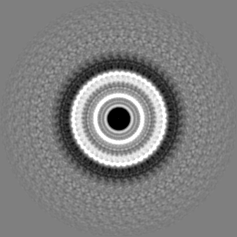
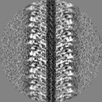
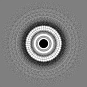
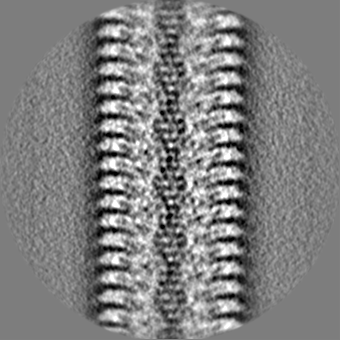
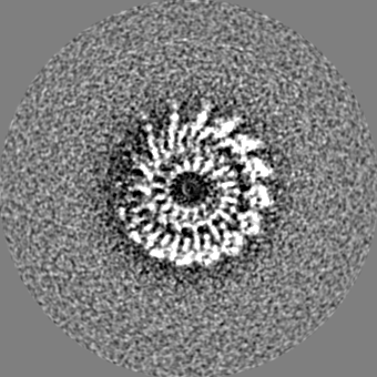
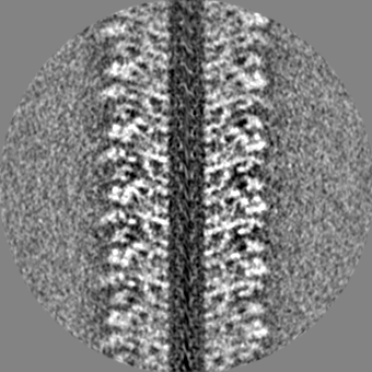
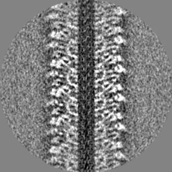
| タイトル | Low-dose cryo-electron ptychographic reconstruction of TMV recorded with CSA of 6.1 mrad | |||||||||
 マップデータ マップデータ | ||||||||||
 試料 試料 |
| |||||||||
 キーワード キーワード | Capsid / Tobacco Mosaic Virus / VIRUS | |||||||||
| 生物種 |  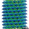 Tobacco mosaic virus (ウイルス) Tobacco mosaic virus (ウイルス) | |||||||||
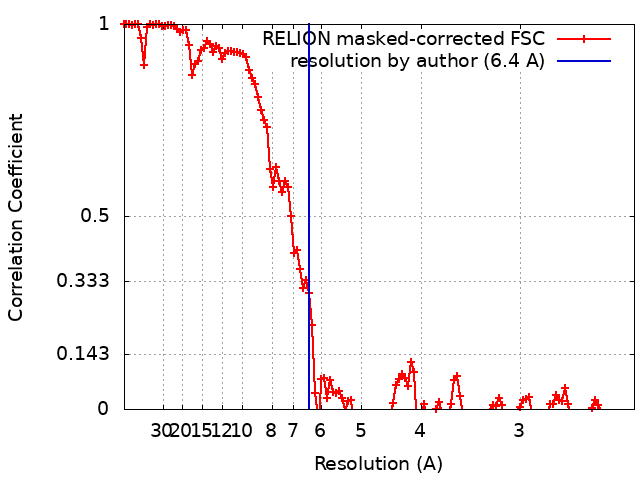
| 手法 | らせん対称体再構成法 / クライオ電子顕微鏡法 / 解像度: 6.4 Å | |||||||||
 データ登録者 データ登録者 | Mohammed I / Stalhberg H / Kucukoglu B | |||||||||
| 資金援助 |  スイス, 1件 スイス, 1件
| |||||||||
 引用 引用 | ジャーナル: Nat Commun / 年: 2024 タイトル: Low-dose cryo-electron ptychography of proteins at sub-nanometer resolution. 著者: Berk Küçükoğlu / Inayathulla Mohammed / Ricardo C Guerrero-Ferreira / Stephanie M Ribet / Georgios Varnavides / Max Leo Leidl / Kelvin Lau / Sergey Nazarov / Alexander Myasnikov / Massimo ...著者: Berk Küçükoğlu / Inayathulla Mohammed / Ricardo C Guerrero-Ferreira / Stephanie M Ribet / Georgios Varnavides / Max Leo Leidl / Kelvin Lau / Sergey Nazarov / Alexander Myasnikov / Massimo Kube / Julika Radecke / Carsten Sachse / Knut Müller-Caspary / Colin Ophus / Henning Stahlberg /   要旨: Cryo-transmission electron microscopy (cryo-EM) of frozen hydrated specimens is an efficient method for the structural analysis of purified biological molecules. However, cryo-EM and cryo-electron ...Cryo-transmission electron microscopy (cryo-EM) of frozen hydrated specimens is an efficient method for the structural analysis of purified biological molecules. However, cryo-EM and cryo-electron tomography are limited by the low signal-to-noise ratio (SNR) of recorded images, making detection of smaller particles challenging. For dose-resilient samples often studied in the physical sciences, electron ptychography - a coherent diffractive imaging technique using 4D scanning transmission electron microscopy (4D-STEM) - has recently demonstrated excellent SNR and resolution down to tens of picometers for thin specimens imaged at room temperature. Here we apply 4D-STEM and ptychographic data analysis to frozen hydrated proteins, reaching sub-nanometer resolution 3D reconstructions. We employ low-dose cryo-EM with an aberration-corrected, convergent electron beam to collect 4D-STEM data for our reconstructions. The high frame rate of the electron detector allows us to record large datasets of electron diffraction patterns with substantial overlaps between the interaction volumes of adjacent scan positions, from which the scattering potentials of the samples are iteratively reconstructed. The reconstructed micrographs show strong SNR enabling the reconstruction of the structure of apoferritin protein at up to 5.8 Å resolution. We also show structural analysis of the Phi92 capsid and sheath, tobacco mosaic virus, and bacteriorhodopsin at slightly lower resolutions. #1: ジャーナル: To Be Publishedタイトル: Low-dose cryo-electron ptychography of proteins at sub-nanometer resolution 著者: Mohammed I / Stalhberg H / Kucukoglu B | |||||||||
| 履歴 |
|
- 構造の表示
構造の表示
| 添付画像 |
|---|

- ダウンロードとリンク
-EMDBアーカイブ
| マップデータ | emd_19885.map.gz | 15.8 MB |  EMDBマップデータ形式 EMDBマップデータ形式 | |
|---|---|---|---|---|
| ヘッダ (付随情報) | emd-19885-v30.xmlemd-19885.xml | 14.6 KB 14.6 KB | 表示 表示 | EMDBヘッダ |
| FSC (解像度算出) | emd_19885_fsc.xml | 12.1 KB | 表示 | FSCデータファイル |
| 画像 |  emd_19885.png emd_19885.png | 73 KB | ||
| マスクデータ | emd_19885_msk_1.map | 149.9 MB | マスクマップ | |
| Filedesc metadata | emd-19885.cif.gz | 4.4 KB | ||
| その他 | emd_19885_half_map_1.map.gzemd_19885_half_map_2.map.gz | 118.7 MB 118.5 MB | ||
| アーカイブディレクトリ |  http://ftp.pdbj.org/pub/emdb/structures/EMD-19885ftp://ftp.pdbj.org/pub/emdb/structures/EMD-19885 http://ftp.pdbj.org/pub/emdb/structures/EMD-19885ftp://ftp.pdbj.org/pub/emdb/structures/EMD-19885 | HTTPS FTP |
-検証レポート
| 文書・要旨 | emd_19885_validation.pdf.gz | 894.6 KB | 表示 | EMDB検証レポート |
|---|---|---|---|---|
| 文書・詳細版 | emd_19885_full_validation.pdf.gz | 894.1 KB | 表示 | |
| XML形式データ | emd_19885_validation.xml.gz | 19.7 KB | 表示 | |
| CIF形式データ | emd_19885_validation.cif.gz | 25.8 KB | 表示 | |
| アーカイブディレクトリ | https://ftp.pdbj.org/pub/emdb/validation_reports/EMD-19885ftp://ftp.pdbj.org/pub/emdb/validation_reports/EMD-19885 | HTTPS FTP |
-関連構造データ


-リンク
| EMDBのページ | EMDB (EBI/PDBe) / EMDataResource |
|---|---|
| 「今月の分子」の関連する項目 |

-マップ
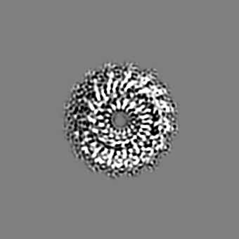
| ファイル | ダウンロード / ファイル: emd_19885.map.gz / 形式: CCP4 / 大きさ: 149.9 MB / タイプ: IMAGE STORED AS FLOATING POINT NUMBER (4 BYTES) | ||||||||||||||||||||||||||||||||||||
|---|---|---|---|---|---|---|---|---|---|---|---|---|---|---|---|---|---|---|---|---|---|---|---|---|---|---|---|---|---|---|---|---|---|---|---|---|---|
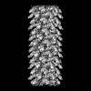
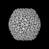
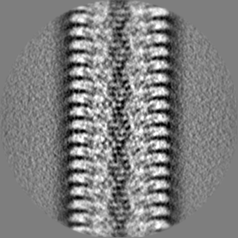
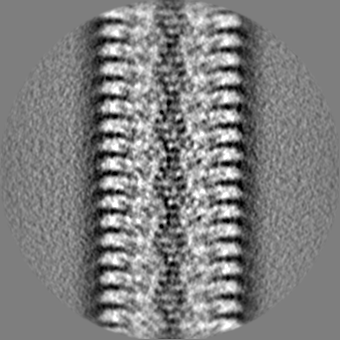
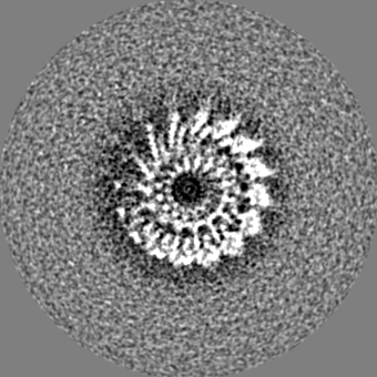
| 投影像・断面図 | 画像のコントロール
画像は Spider により作成 | ||||||||||||||||||||||||||||||||||||
| ボクセルのサイズ | X=Y=Z: 1.1659 Å | ||||||||||||||||||||||||||||||||||||
| 密度 |
| ||||||||||||||||||||||||||||||||||||
| 対称性 | 空間群: 1 | ||||||||||||||||||||||||||||||||||||
| 詳細 | EMDB XML:
|
 Z (Sec.)
 Y (Row.)
 X (Col.)


-添付データ
-マスク #1
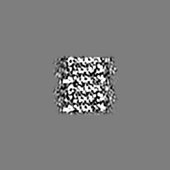
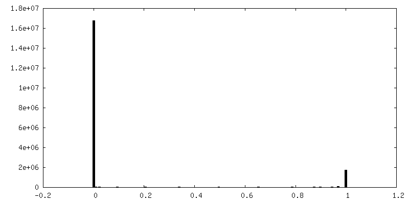
| ファイル | emd_19885_msk_1.map | ||||||||||||
|---|---|---|---|---|---|---|---|---|---|---|---|---|---|
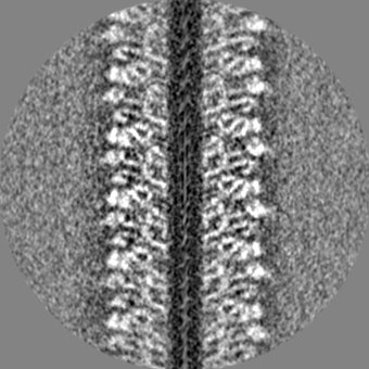
| 投影像・断面図 |
| ||||||||||||
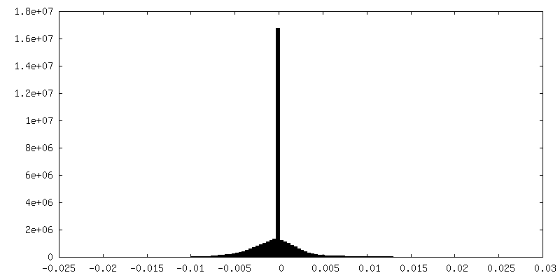
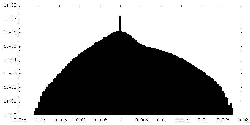
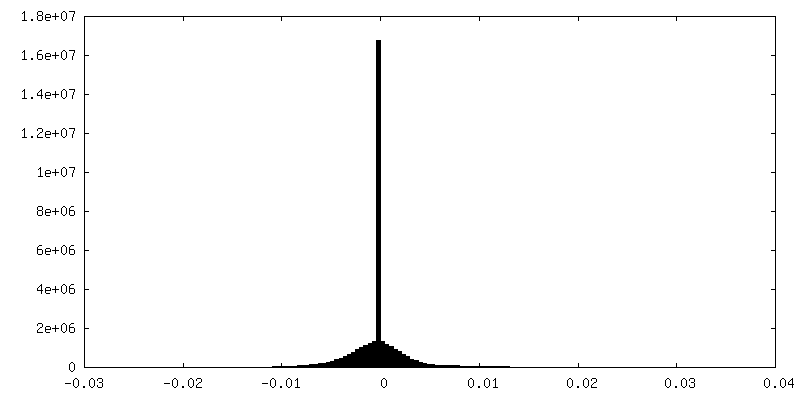
| 密度ヒストグラム |


-ハーフマップ: #1
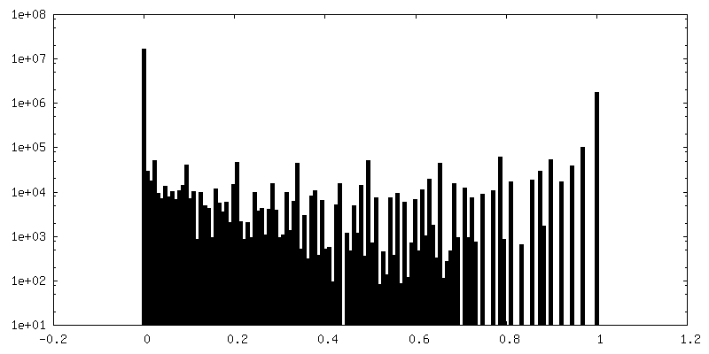
| ファイル | emd_19885_half_map_1.map | ||||||||||||
|---|---|---|---|---|---|---|---|---|---|---|---|---|---|
| 投影像・断面図 |
| ||||||||||||
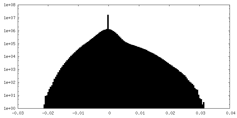
| 密度ヒストグラム |
-ハーフマップ: #2
| ファイル | emd_19885_half_map_2.map | ||||||||||||
|---|---|---|---|---|---|---|---|---|---|---|---|---|---|
| 投影像・断面図 |
| ||||||||||||
| 密度ヒストグラム |

- 試料の構成要素
-全体 : Tobacco mosaic virus
| 全体 | 名称: Tobacco mosaic virus (ウイルス) |
|---|---|
| 要素 |
|
-超分子 #1: Tobacco mosaic virus
| 超分子 | 名称: Tobacco mosaic virus / タイプ: virus / ID: 1 / 親要素: 0 / NCBI-ID: 12242 / 生物種: Tobacco mosaic virus / ウイルスタイプ: VIRION / ウイルス・単離状態: OTHER / ウイルス・エンベロープ: Yes / ウイルス・中空状態: No |
|---|---|
| 分子量 | 理論値: 40.0 MDa |
-実験情報
-構造解析
| 手法 | クライオ電子顕微鏡法 |
|---|---|
 解析 解析 | らせん対称体再構成法 |
| 試料の集合状態 | filament |
-試料調製
| 緩衝液 | pH: 7.4 |
|---|---|
| グリッド | モデル: Quantifoil R1.2/1.3 / 材質: GOLD / メッシュ: 300 / 前処理 - タイプ: GLOW DISCHARGE / 前処理 - 時間: 60 sec. |
| 凍結 | 凍結剤: ETHANE / チャンバー内湿度: 95 % / チャンバー内温度: 283 K / 装置: FEI VITROBOT MARK IV |
- 電子顕微鏡法
| 顕微鏡 | FEI TITAN KRIOS |
|---|---|
| 撮影 | フィルム・検出器のモデル: DECTRIS ELA (1k x 0.5k) 平均電子線量: 32.0 e/Å2 |
| 電子線 | 加速電圧: 300 kV / 電子線源:  FIELD EMISSION GUN FIELD EMISSION GUN |
| 電子光学系 | 照射モード: SPOT SCAN / 撮影モード: DIFFRACTION / Cs: 0.1 mm / 最大 デフォーカス(公称値): 1.8 µm 最小 デフォーカス(公称値): 1.4000000000000001 µm |
| 実験機器 |  モデル: Titan Krios / 画像提供: FEI Company |