 ムービー
ムービー コントローラー


+ データを開く

- 基本情報
基本情報
| 登録情報 |  | |||||||||
|---|---|---|---|---|---|---|---|---|---|---|
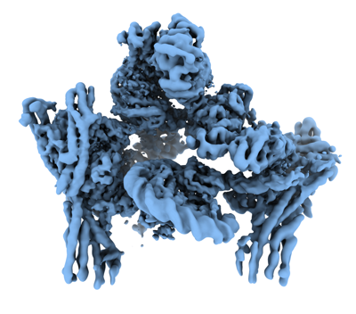
| タイトル | Flexible reconstruction of the yeast inner kinetochore bound to a CENP-A nucleosome (EMPIAR-11890) | |||||||||
 マップデータ マップデータ | local resolution filtered map from the DynaMight half maps | |||||||||
 試料 試料 |
| |||||||||
 キーワード キーワード | kinetochore / point centromere / CENP-A nucleosome / topological entrapment / centromeric DNA / CELL CYCLE | |||||||||
| 生物種 |  | |||||||||
| 手法 | 単粒子再構成法 / クライオ電子顕微鏡法 / 解像度: 4.47 Å | |||||||||
 データ登録者 データ登録者 | Schwab J / Kimanius D / Burt A / Dendooven T / Scheres SHW | |||||||||
| 資金援助 |  英国, 1件 英国, 1件
| |||||||||
 引用 引用 | ジャーナル: Sci Adv / 年: 2023 タイトル: Cryo-EM structure of the complete inner kinetochore of the budding yeast point centromere. 著者: Tom Dendooven / Ziguo Zhang / Jing Yang / Stephen H McLaughlin / Johannes Schwab / Sjors H W Scheres / Stanislau Yatskevich / David Barford / 要旨: The point centromere of budding yeast specifies assembly of the large kinetochore complex to mediate chromatid segregation. Kinetochores comprise the centromere-associated inner kinetochore (CCAN) ...The point centromere of budding yeast specifies assembly of the large kinetochore complex to mediate chromatid segregation. Kinetochores comprise the centromere-associated inner kinetochore (CCAN) complex and the microtubule-binding outer kinetochore KNL1-MIS12-NDC80 (KMN) network. The budding yeast inner kinetochore also contains the DNA binding centromere-binding factor 1 (CBF1) and CBF3 complexes. We determined the cryo-electron microscopy structure of the yeast inner kinetochore assembled onto the centromere-specific centromere protein A nucleosomes (CENP-A). This revealed a central CENP-A with extensively unwrapped DNA ends. These free DNA duplexes bind two CCAN protomers, one of which entraps DNA topologically, positioned on the centromere DNA element I (CDEI) motif by CBF1. The two CCAN protomers are linked through CBF3 forming an arch-like configuration. With a structural mechanism for how CENP-A can also be linked to KMN involving only CENP-QU, we present a model for inner kinetochore assembly onto a point centromere and how it organizes the outer kinetochore for chromosome attachment to the mitotic spindle. #1: ジャーナル: biorxivタイトル: DynaMight: estimating molecular motions with improved reconstruction from cryo-EM images 著者: Schwab J / Kimanius D / Burt A / Dendooven T / Scheres SHW | |||||||||
| 履歴 |
|
- 構造の表示
構造の表示
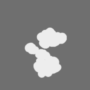
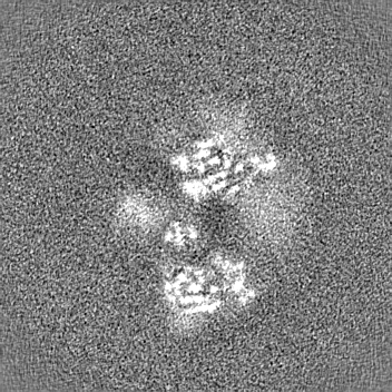
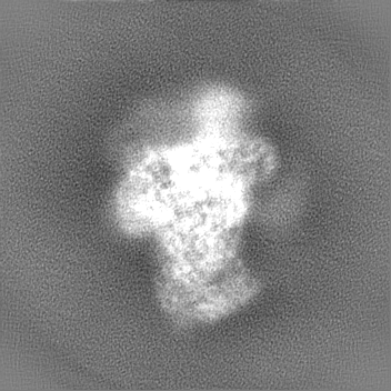
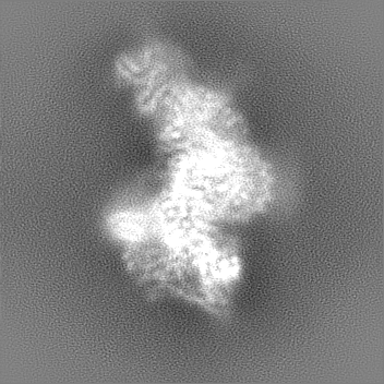
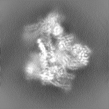
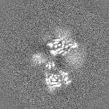
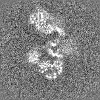
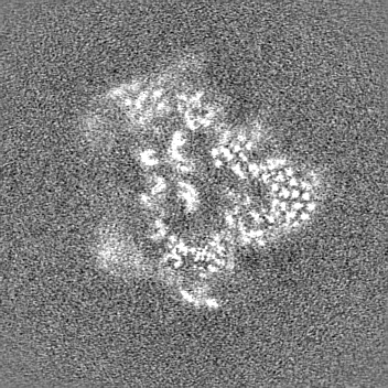
| 添付画像 |
|---|
- ダウンロードとリンク
-EMDBアーカイブ
| マップデータ | emd_19794.map.gz | 96.6 MB |  EMDBマップデータ形式 EMDBマップデータ形式 | |
|---|---|---|---|---|
| ヘッダ (付随情報) | emd-19794-v30.xmlemd-19794.xml | 14.3 KB 14.3 KB | 表示 表示 | EMDBヘッダ |
| 画像 |  emd_19794.png emd_19794.png | 125 KB | ||
| マスクデータ | emd_19794_msk_1.map | 166.4 MB | マスクマップ | |
| Filedesc metadata | emd-19794.cif.gz | 4.1 KB | ||
| その他 | emd_19794_half_map_1.map.gzemd_19794_half_map_2.map.gz | 154.6 MB 154.7 MB | ||
| アーカイブディレクトリ |  http://ftp.pdbj.org/pub/emdb/structures/EMD-19794ftp://ftp.pdbj.org/pub/emdb/structures/EMD-19794 http://ftp.pdbj.org/pub/emdb/structures/EMD-19794ftp://ftp.pdbj.org/pub/emdb/structures/EMD-19794 | HTTPS FTP |
-検証レポート
| 文書・要旨 | emd_19794_validation.pdf.gz | 1002.9 KB | 表示 | EMDB検証レポート |
|---|---|---|---|---|
| 文書・詳細版 | emd_19794_full_validation.pdf.gz | 1002.4 KB | 表示 | |
| XML形式データ | emd_19794_validation.xml.gz | 14.2 KB | 表示 | |
| CIF形式データ | emd_19794_validation.cif.gz | 16.8 KB | 表示 | |
| アーカイブディレクトリ | https://ftp.pdbj.org/pub/emdb/validation_reports/EMD-19794ftp://ftp.pdbj.org/pub/emdb/validation_reports/EMD-19794 | HTTPS FTP |
-関連構造データ


-リンク
| EMDBのページ | EMDB (EBI/PDBe) / EMDataResource |
|---|
-マップ
| ファイル | ダウンロード / ファイル: emd_19794.map.gz / 形式: CCP4 / 大きさ: 166.4 MB / タイプ: IMAGE STORED AS FLOATING POINT NUMBER (4 BYTES) | ||||||||||||||||||||||||||||||||||||
|---|---|---|---|---|---|---|---|---|---|---|---|---|---|---|---|---|---|---|---|---|---|---|---|---|---|---|---|---|---|---|---|---|---|---|---|---|---|
| 注釈 | local resolution filtered map from the DynaMight half maps | ||||||||||||||||||||||||||||||||||||
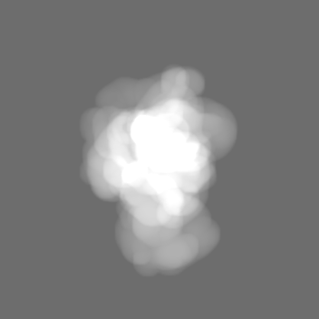
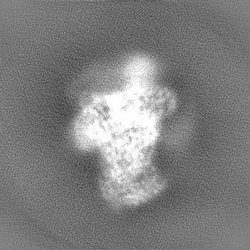
| 投影像・断面図 | 画像のコントロール
画像は Spider により作成 | ||||||||||||||||||||||||||||||||||||
| ボクセルのサイズ | X=Y=Z: 1.08 Å | ||||||||||||||||||||||||||||||||||||
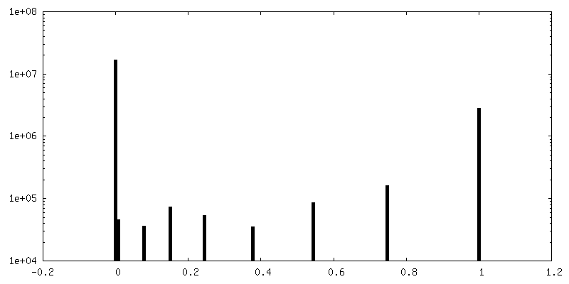
| 密度 |
| ||||||||||||||||||||||||||||||||||||
| 対称性 | 空間群: 1 | ||||||||||||||||||||||||||||||||||||
| 詳細 | EMDB XML:
|
 Z (Sec.)
 Y (Row.)
 X (Col.)


-添付データ
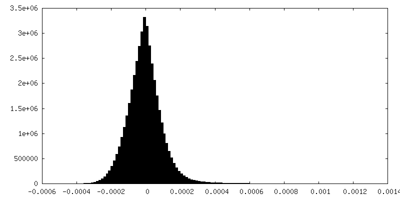
-マスク #1
| ファイル | emd_19794_msk_1.map | ||||||||||||
|---|---|---|---|---|---|---|---|---|---|---|---|---|---|
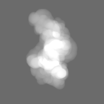
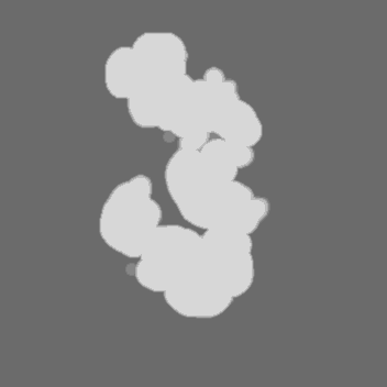
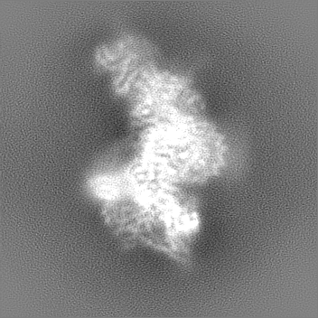
| 投影像・断面図 |
| ||||||||||||
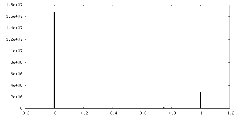
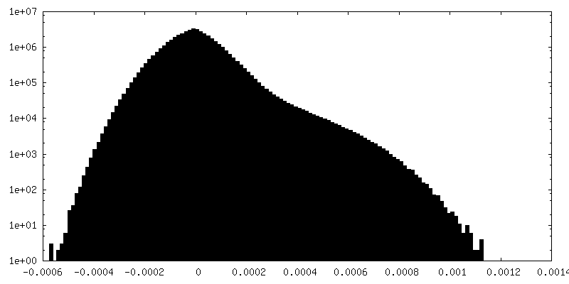
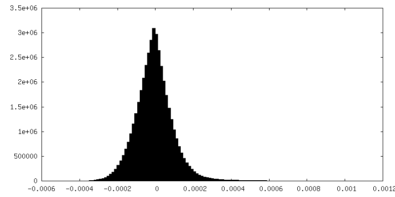
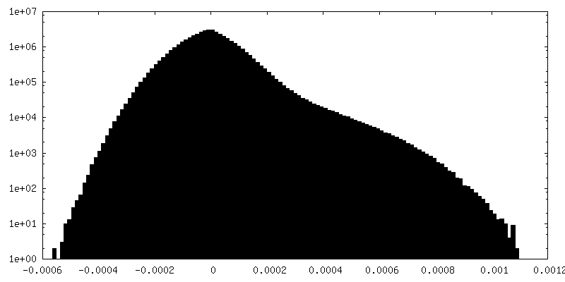
| 密度ヒストグラム |
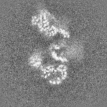
-ハーフマップ: unfiltered, unmasked half map from reconstruction with DynaMight
| ファイル | emd_19794_half_map_1.map | ||||||||||||
|---|---|---|---|---|---|---|---|---|---|---|---|---|---|
| 注釈 | unfiltered, unmasked half map from reconstruction with DynaMight | ||||||||||||
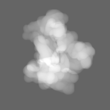
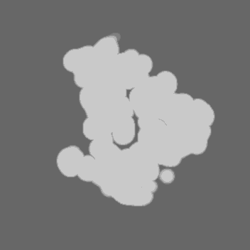
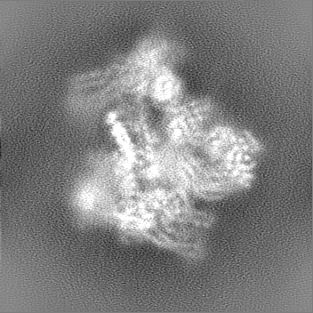
| 投影像・断面図 |
| ||||||||||||
| 密度ヒストグラム |
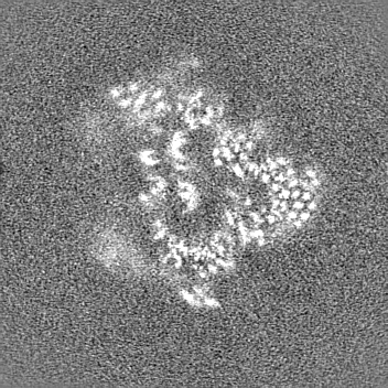
-ハーフマップ: unfiltered, unmasked half map from reconstruction with DynaMight
| ファイル | emd_19794_half_map_2.map | ||||||||||||
|---|---|---|---|---|---|---|---|---|---|---|---|---|---|
| 注釈 | unfiltered, unmasked half map from reconstruction with DynaMight | ||||||||||||
| 投影像・断面図 |
| ||||||||||||
| 密度ヒストグラム |
- 試料の構成要素
-全体 : A complex of CBF1-CCAN bound to centromeric C0N3 DNA
| 全体 | 名称: A complex of CBF1-CCAN bound to centromeric C0N3 DNA |
|---|---|
| 要素 |
|
-超分子 #1: A complex of CBF1-CCAN bound to centromeric C0N3 DNA
| 超分子 | 名称: A complex of CBF1-CCAN bound to centromeric C0N3 DNA タイプ: complex / ID: 1 / 親要素: 0 |
|---|---|
| 由来(天然) | 生物種: |
-実験情報
-構造解析
| 手法 | クライオ電子顕微鏡法 |
|---|---|
 解析 解析 | 単粒子再構成法 |
| 試料の集合状態 | particle |
-試料調製
| 緩衝液 | pH: 8 |
|---|---|
| 凍結 | 凍結剤: ETHANE |
- 電子顕微鏡法
| 顕微鏡 | FEI TITAN KRIOS |
|---|---|
| 撮影 | フィルム・検出器のモデル: GATAN K3 BIOQUANTUM (6k x 4k) 平均電子線量: 40.0 e/Å2 |
| 電子線 | 加速電圧: 300 kV / 電子線源:  FIELD EMISSION GUN FIELD EMISSION GUN |
| 電子光学系 | 照射モード: SPOT SCAN / 撮影モード: BRIGHT FIELD / 最大 デフォーカス(公称値): 2.6 µm / 最小 デフォーカス(公称値): 1.0 µm |
| 実験機器 |  モデル: Titan Krios / 画像提供: FEI Company |
-画像解析
| 初期モデル | モデルのタイプ: NONE |
|---|---|
| 最終 再構成 | 解像度のタイプ: BY AUTHOR / 解像度: 4.47 Å / 解像度の算出法: FSC 0.143 CUT-OFF / 使用した粒子像数: 108672 |
| 初期 角度割当 | タイプ: MAXIMUM LIKELIHOOD |
| 最終 角度割当 | タイプ: MAXIMUM LIKELIHOOD |
