 Movie
Movie Controller

[English] 日本語
 Yorodumi
- EMDB-19791: Flexible reconstruction of a pre-catalytic spliceosome (EMPIAR-10... -
+ Open data

- Basic information
| Entry |  | |||||||||
|---|---|---|---|---|---|---|---|---|---|---|
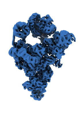
| Title | Flexible reconstruction of a pre-catalytic spliceosome (EMPIAR-10180) using DynaMight | |||||||||
 Map data Map data | local resolution filtered map from the DynaMight half maps | |||||||||
 Sample Sample |
| |||||||||
 Keywords Keywords | Spliceosome / SPLICING | |||||||||
| Biological species |  | |||||||||
| Method | single particle reconstruction / cryo EM / Resolution: 5.98 Å | |||||||||
 Authors Authors | Schwab J / Scheres SHW | |||||||||
| Funding support |  United Kingdom, 1 items United Kingdom, 1 items
| |||||||||
 Citation Citation | Journal: Nature / Year: 2017 Title: Structure of a pre-catalytic spliceosome. Authors: Clemens Plaschka / Pei-Chun Lin / Kiyoshi Nagai / Abstract: Intron removal requires assembly of the spliceosome on precursor mRNA (pre-mRNA) and extensive remodelling to form the spliceosome's catalytic centre. Here we report the cryo-electron microscopy ...Intron removal requires assembly of the spliceosome on precursor mRNA (pre-mRNA) and extensive remodelling to form the spliceosome's catalytic centre. Here we report the cryo-electron microscopy structure of the yeast Saccharomyces cerevisiae pre-catalytic B complex spliceosome at near-atomic resolution. The mobile U2 small nuclear ribonucleoprotein particle (snRNP) associates with U4/U6.U5 tri-snRNP through the U2/U6 helix II and an interface between U4/U6 di-snRNP and the U2 snRNP SF3b-containing domain, which also transiently contacts the helicase Brr2. The 3' region of the U2 snRNP is flexibly attached to the SF3b-containing domain and protrudes over the concave surface of tri-snRNP, where the U1 snRNP may reside before its release from the pre-mRNA 5' splice site. The U6 ACAGAGA sequence forms a hairpin that weakly tethers the 5' splice site. The B complex proteins Prp38, Snu23 and Spp381 bind the Prp8 N-terminal domain and stabilize U6 ACAGAGA stem-pre-mRNA and Brr2-U4 small nuclear RNA interactions. These results provide important insights into the events leading to active site formation. #1: Journal: BiorxivTitle: DynaMight: estimating molecular motions with improved reconstruction from cryo-EM images Authors: Schwab J / Kimanius D / Burt A / Dendooven T / Scheres SHW | |||||||||
| History |
|
- Structure visualization
| Supplemental images |
|---|
- Downloads & links
-EMDB archive
| Map data | emd_19791.map.gz | 70.6 MB |  EMDB map data format EMDB map data format | |
|---|---|---|---|---|
| Header (meta data) | emd-19791-v30.xmlemd-19791.xml | 13.9 KB 13.9 KB | Display Display | EMDB header |
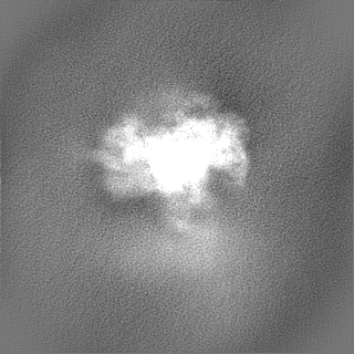
| Images |  emd_19791.png emd_19791.png | 79.6 KB | ||
| Filedesc metadata | emd-19791.cif.gz | 4 KB | ||
| Others | emd_19791_half_map_1.map.gzemd_19791_half_map_2.map.gz | 116 MB 116 MB | ||
| Archive directory |  http://ftp.pdbj.org/pub/emdb/structures/EMD-19791ftp://ftp.pdbj.org/pub/emdb/structures/EMD-19791 http://ftp.pdbj.org/pub/emdb/structures/EMD-19791ftp://ftp.pdbj.org/pub/emdb/structures/EMD-19791 | HTTPS FTP |
-Related structure data


-Links
| EMDB pages | EMDB (EBI/PDBe) / EMDataResource |
|---|
-Map
| File | Download / File: emd_19791.map.gz / Format: CCP4 / Size: 125 MB / Type: IMAGE STORED AS FLOATING POINT NUMBER (4 BYTES) | ||||||||||||||||||||||||||||||||||||
|---|---|---|---|---|---|---|---|---|---|---|---|---|---|---|---|---|---|---|---|---|---|---|---|---|---|---|---|---|---|---|---|---|---|---|---|---|---|
| Annotation | local resolution filtered map from the DynaMight half maps | ||||||||||||||||||||||||||||||||||||
| Projections & slices | Image control
Images are generated by Spider. | ||||||||||||||||||||||||||||||||||||
| Voxel size | X=Y=Z: 1.7 Å | ||||||||||||||||||||||||||||||||||||
| Density |
| ||||||||||||||||||||||||||||||||||||
| Symmetry | Space group: 1 | ||||||||||||||||||||||||||||||||||||
| Details | EMDB XML:
|
 Z (Sec.)
 Y (Row.)
 X (Col.)


-Supplemental data
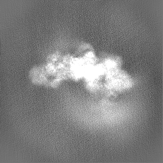
-Half map: unfiltered, unmasked half map reconstructed with DynaMight
| File | emd_19791_half_map_1.map | ||||||||||||
|---|---|---|---|---|---|---|---|---|---|---|---|---|---|
| Annotation | unfiltered, unmasked half map reconstructed with DynaMight | ||||||||||||
| Projections & Slices |
| ||||||||||||
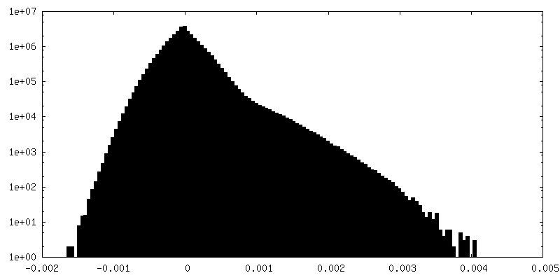
| Density Histograms |


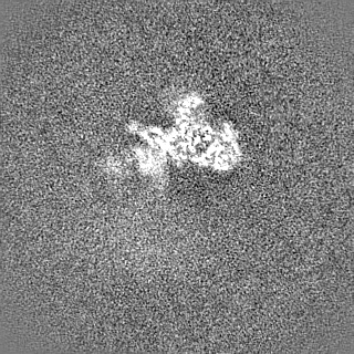


-Half map: unfiltered, unmasked half map reconstructed with DynaMight
| File | emd_19791_half_map_2.map | ||||||||||||
|---|---|---|---|---|---|---|---|---|---|---|---|---|---|
| Annotation | unfiltered, unmasked half map reconstructed with DynaMight | ||||||||||||
| Projections & Slices |
| ||||||||||||
| Density Histograms |


- Sample components
-Entire : Pre-catalytic B complex Spliceosome
| Entire | Name: Pre-catalytic B complex Spliceosome |
|---|---|
| Components |
|
-Supramolecule #1: Pre-catalytic B complex Spliceosome
| Supramolecule | Name: Pre-catalytic B complex Spliceosome / type: complex / ID: 1 / Parent: 0 |
|---|---|
| Source (natural) | Organism: |
-Experimental details
-Structure determination
| Method | cryo EM |
|---|---|
 Processing Processing | single particle reconstruction |
| Aggregation state | particle |
-Sample preparation
| Buffer | pH: 7.9 |
|---|---|
| Vitrification | Cryogen name: ETHANE |
- Electron microscopy
| Microscope | FEI TITAN KRIOS |
|---|---|
| Image recording | Film or detector model: GATAN K2 SUMMIT (4k x 4k) / Average electron dose: 56.0 e/Å2 |
| Electron beam | Acceleration voltage: 300 kV / Electron source:  FIELD EMISSION GUN FIELD EMISSION GUN |
| Electron optics | Illumination mode: FLOOD BEAM / Imaging mode: BRIGHT FIELD / Nominal defocus max: 5.3 µm / Nominal defocus min: 0.35000000000000003 µm |
| Experimental equipment |  Model: Titan Krios / Image courtesy: FEI Company |
-Image processing
| Startup model | Type of model: OTHER |
|---|---|
| Final reconstruction | Resolution.type: BY AUTHOR / Resolution: 5.98 Å / Resolution method: FSC 0.143 CUT-OFF / Number images used: 44537 |
| Initial angle assignment | Type: PROJECTION MATCHING |
| Final angle assignment | Type: MAXIMUM LIKELIHOOD / Software - Name: RELION (ver. 5.0) |
