ムービー
ムービー コントローラー


+ データを開く

- 基本情報
基本情報
| 登録情報 |  | |||||||||
|---|---|---|---|---|---|---|---|---|---|---|
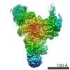
| タイトル | Flexible reconstruction of the yeast U4/U6.U5 tri-snRNP (EMPIAR-10073) using DynaMight | |||||||||
 マップデータ マップデータ | local resolution filtered map from the DynaMight half maps. Reconstruction was done with a subset of 86,624 particles of EMPIAR-10073 with deformation correction | |||||||||
 試料 試料 |
| |||||||||
 キーワード キーワード | Spliceosome / SPLICING | |||||||||
| 生物種 |  | |||||||||
| 手法 | 単粒子再構成法 / クライオ電子顕微鏡法 / 解像度: 3.91 Å | |||||||||
 データ登録者 データ登録者 | Schwab J / Scheres SHW | |||||||||
| 資金援助 |  英国, 1件 英国, 1件
| |||||||||
 引用 引用 | ジャーナル: Nature / 年: 2016 タイトル: Cryo-EM structure of the yeast U4/U6.U5 tri-snRNP at 3.7 Å resolution. 著者: Thi Hoang Duong Nguyen / Wojciech P Galej / Xiao-Chen Bai / Chris Oubridge / Andrew J Newman / Sjors H W Scheres / Kiyoshi Nagai / 要旨: U4/U6.U5 tri-snRNP represents a substantial part of the spliceosome before activation. A cryo-electron microscopy structure of Saccharomyces cerevisiae U4/U6.U5 tri-snRNP at 3.7 Å resolution led ...U4/U6.U5 tri-snRNP represents a substantial part of the spliceosome before activation. A cryo-electron microscopy structure of Saccharomyces cerevisiae U4/U6.U5 tri-snRNP at 3.7 Å resolution led to an essentially complete atomic model comprising 30 proteins plus U4/U6 and U5 small nuclear RNAs (snRNAs). The structure reveals striking interweaving interactions of the protein and RNA components, including extended polypeptides penetrating into subunit interfaces. The invariant ACAGAGA sequence of U6 snRNA, which base-pairs with the 5'-splice site during catalytic activation, forms a hairpin stabilized by Dib1 and Prp8 while the adjacent nucleotides interact with the exon binding loop 1 of U5 snRNA. Snu114 harbours GTP, but its putative catalytic histidine is held away from the γ-phosphate by hydrogen bonding to a tyrosine in the amino-terminal domain of Prp8. Mutation of this histidine to alanine has no detectable effect on yeast growth. The structure provides important new insights into the spliceosome activation process leading to the formation of the catalytic centre. #1: ジャーナル: Biorxivタイトル: DynaMight: estimating molecular motions with improved reconstruction from cryo-EM images 著者: Schwab J / Kimanius D / Burt A / Dendooven T / Scheres SHW | |||||||||
| 履歴 |
|
- 構造の表示
構造の表示
| 添付画像 |
|---|

- ダウンロードとリンク
-EMDBアーカイブ
| マップデータ | emd_19789.map.gz | 5.2 MB |  EMDBマップデータ形式 EMDBマップデータ形式 | |
|---|---|---|---|---|
| ヘッダ (付随情報) | emd-19789-v30.xmlemd-19789.xml | 14.8 KB 14.8 KB | 表示 表示 | EMDBヘッダ |
| 画像 |  emd_19789.png emd_19789.png | 128.7 KB | ||
| Filedesc metadata | emd-19789.cif.gz | 4.1 KB | ||
| その他 | emd_19789_half_map_1.map.gzemd_19789_half_map_2.map.gz | 194.7 MB 194.7 MB | ||
| アーカイブディレクトリ |  http://ftp.pdbj.org/pub/emdb/structures/EMD-19789ftp://ftp.pdbj.org/pub/emdb/structures/EMD-19789 http://ftp.pdbj.org/pub/emdb/structures/EMD-19789ftp://ftp.pdbj.org/pub/emdb/structures/EMD-19789 | HTTPS FTP |
-検証レポート
| 文書・要旨 | emd_19789_validation.pdf.gz | 815.7 KB | 表示 | EMDB検証レポート |
|---|---|---|---|---|
| 文書・詳細版 | emd_19789_full_validation.pdf.gz | 815.3 KB | 表示 | |
| XML形式データ | emd_19789_validation.xml.gz | 15 KB | 表示 | |
| CIF形式データ | emd_19789_validation.cif.gz | 18.1 KB | 表示 | |
| アーカイブディレクトリ | https://ftp.pdbj.org/pub/emdb/validation_reports/EMD-19789ftp://ftp.pdbj.org/pub/emdb/validation_reports/EMD-19789 | HTTPS FTP |
-関連構造データ


-リンク
| EMDBのページ | EMDB (EBI/PDBe) / EMDataResource |
|---|
-マップ
| ファイル | ダウンロード / ファイル: emd_19789.map.gz / 形式: CCP4 / 大きさ: 209.3 MB / タイプ: IMAGE STORED AS FLOATING POINT NUMBER (4 BYTES) | ||||||||||||||||||||||||||||||||||||
|---|---|---|---|---|---|---|---|---|---|---|---|---|---|---|---|---|---|---|---|---|---|---|---|---|---|---|---|---|---|---|---|---|---|---|---|---|---|
| 注釈 | local resolution filtered map from the DynaMight half maps. Reconstruction was done with a subset of 86,624 particles of EMPIAR-10073 with deformation correction | ||||||||||||||||||||||||||||||||||||
| 投影像・断面図 | 画像のコントロール
画像は Spider により作成 | ||||||||||||||||||||||||||||||||||||
| ボクセルのサイズ | X=Y=Z: 1.40001 Å | ||||||||||||||||||||||||||||||||||||
| 密度 |
| ||||||||||||||||||||||||||||||||||||
| 対称性 | 空間群: 1 | ||||||||||||||||||||||||||||||||||||
| 詳細 | EMDB XML:
|
 Z (Sec.)
 Y (Row.)
 X (Col.)


-添付データ
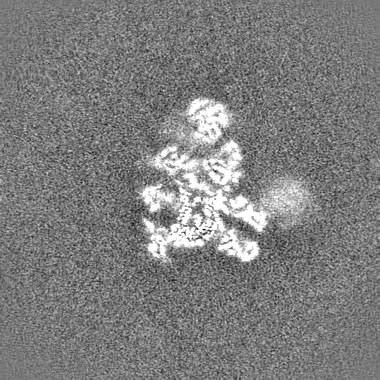
-ハーフマップ: Unfiltered half map obtained with DynaMight reconstruction
| ファイル | emd_19789_half_map_1.map | ||||||||||||
|---|---|---|---|---|---|---|---|---|---|---|---|---|---|
| 注釈 | Unfiltered half map obtained with DynaMight reconstruction | ||||||||||||
| 投影像・断面図 |
| ||||||||||||
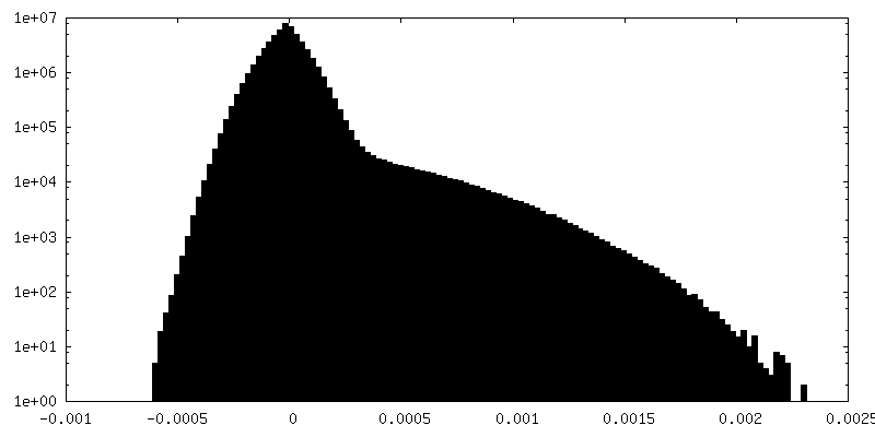
| 密度ヒストグラム |


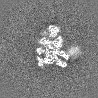
-ハーフマップ: Unfiltered half map obtained with DynaMight reconstruction
| ファイル | emd_19789_half_map_2.map | ||||||||||||
|---|---|---|---|---|---|---|---|---|---|---|---|---|---|
| 注釈 | Unfiltered half map obtained with DynaMight reconstruction | ||||||||||||
| 投影像・断面図 |
| ||||||||||||
| 密度ヒストグラム |


- 試料の構成要素
-全体 : The overall structure of the yeast spliceosomal U4/U6.U5 tri-snRNP
| 全体 | 名称: The overall structure of the yeast spliceosomal U4/U6.U5 tri-snRNP |
|---|---|
| 要素 |
|
-超分子 #1: The overall structure of the yeast spliceosomal U4/U6.U5 tri-snRNP
| 超分子 | 名称: The overall structure of the yeast spliceosomal U4/U6.U5 tri-snRNP タイプ: complex / ID: 1 / 親要素: 0 |
|---|---|
| 由来(天然) | 生物種: |
-実験情報
-構造解析
| 手法 | クライオ電子顕微鏡法 |
|---|---|
 解析 解析 | 単粒子再構成法 |
| 試料の集合状態 | particle |
-試料調製
| 緩衝液 | pH: 7.9 |
|---|---|
| 凍結 | 凍結剤: NITROGEN |
- 電子顕微鏡法
| 顕微鏡 | FEI TITAN KRIOS |
|---|---|
| 撮影 | フィルム・検出器のモデル: GATAN K2 SUMMIT (4k x 4k) 平均電子線量: 38.0 e/Å2 |
| 電子線 | 加速電圧: 300 kV / 電子線源:  FIELD EMISSION GUN FIELD EMISSION GUN |
| 電子光学系 | 照射モード: FLOOD BEAM / 撮影モード: BRIGHT FIELD / 最大 デフォーカス(公称値): 3.5 µm / 最小 デフォーカス(公称値): 0.5 µm |
| 実験機器 |  モデル: Titan Krios / 画像提供: FEI Company |