ムービー
ムービー コントローラー


+ データを開く

- 基本情報
基本情報
| 登録情報 |  | |||||||||
|---|---|---|---|---|---|---|---|---|---|---|
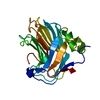
| タイトル | Cryo-EM structure of the C terminal region of PTX3 with a section of coiled-coil | |||||||||
 マップデータ マップデータ | ||||||||||
 試料 試料 |
| |||||||||
 キーワード キーワード | PTX3 / Pentraxin / pattern / recognition / coiled-coil / coil / innate / immunity / extracellular / octamer / Pentraxin-related / cryo-EM / IMMUNE SYSTEM | |||||||||
| 機能・相同性 |  機能・相同性情報 機能・相同性情報(1->3)-beta-D-glucan binding / negative regulation by host of viral glycoprotein metabolic process / negative regulation of glycoprotein metabolic process / ovarian cumulus expansion / host-mediated suppression of viral proces / opsonization / complement component C1q complex binding / response to yeast / host-mediated suppression of symbiont invasion / virion binding ...(1->3)-beta-D-glucan binding / negative regulation by host of viral glycoprotein metabolic process / negative regulation of glycoprotein metabolic process / ovarian cumulus expansion / host-mediated suppression of viral proces / opsonization / complement component C1q complex binding / response to yeast / host-mediated suppression of symbiont invasion / virion binding / positive regulation of phagocytosis / extracellular matrix organization / extracellular matrix / specific granule lumen / positive regulation of nitric oxide biosynthetic process / tertiary granule lumen / inflammatory response / innate immune response / Neutrophil degranulation / extracellular space / extracellular region / identical protein binding 類似検索 - 分子機能 | |||||||||
| 生物種 |  Homo sapiens (ヒト) Homo sapiens (ヒト) | |||||||||
| 手法 | 単粒子再構成法 / クライオ電子顕微鏡法 / 解像度: 3.33 Å | |||||||||
 データ登録者 データ登録者 | Snee M / Shah A / Lockhart-Cairns M / Collins R / Levy C / Baldock C / Day A | |||||||||
| 資金援助 |  英国, 1件 英国, 1件
| |||||||||
 引用 引用 | ジャーナル: Matrix Biol / 年: 2025 タイトル: The structural organisation of pentraxin-3 and its interactions with heavy chains of inter-α-inhibitor regulate crosslinking of the hyaluronan matrix. 著者: Anokhi Shah / Xiaoli Zhang / Matthew Snee / Michael P Lockhart-Cairns / Colin W Levy / Thomas A Jowitt / Holly L Birchenough / Louisa Dean / Richard Collins / Rebecca J Dodd / Abigail R E ...著者: Anokhi Shah / Xiaoli Zhang / Matthew Snee / Michael P Lockhart-Cairns / Colin W Levy / Thomas A Jowitt / Holly L Birchenough / Louisa Dean / Richard Collins / Rebecca J Dodd / Abigail R E Roberts / Jan J Enghild / Alberto Mantovani / Juan Fontana / Clair Baldock / Antonio Inforzato / Ralf P Richter / Anthony J Day /   要旨: Pentraxin-3 (PTX3) is an octameric protein, comprised of eight identical protomers, that has diverse functions in reproductive biology, innate immunity and cancer. PTX3 interacts with the large ...Pentraxin-3 (PTX3) is an octameric protein, comprised of eight identical protomers, that has diverse functions in reproductive biology, innate immunity and cancer. PTX3 interacts with the large polysaccharide hyaluronan (HA) to which heavy chains (HCs) of the inter-α-inhibitor (IαI) family of proteoglycans are covalently attached, playing a key role in the (non-covalent) crosslinking of HC•HA complexes. These interactions stabilise the cumulus matrix, essential for ovulation and fertilisation in mammals, and are also implicated in the formation of pathogenic matrices in the context of viral lung infections. To better understand the physiological and pathological roles of PTX3 we have analysed how its quaternary structure underpins HA crosslinking via its interactions with HCs. A combination of X-ray crystallography, cryo-electron microscopy (cryo-EM) and AlphaFold predictive modelling revealed that the C-terminal pentraxin domains of the PTX3 octamer are arranged in a central cube, with two long extensions on either side, each formed from four protomers assembled into tetrameric coiled-coil regions, essentially as described by (Noone et al., 2022; doi:10.1073/pnas.2208144119). From crystallography and cryo-EM data, we identified a network of inter-protomer salt bridges that facilitate the assembly of the octamer. Small angle X-ray scattering (SAXS) validated our model for the octameric protein, including the analysis of two PTX3 constructs: a tetrameric 'Half-PTX3' and a construct missing the 24 N-terminal residues (Δ1-24_PTX3). SAXS determined a length of ∼520 Å for PTX3 and, combined with 3D variability analysis of cryo-EM data, defined the flexibility of the N-terminal extensions. Biophysical analyses revealed that the prototypical heavy chain HC1 does not interact with PTX3 at pH 7.4, consistent with our previous studies showing that, at this pH, PTX3 only associates with HC•HA complexes if they are formed in its presence. However, PTX3 binds to HC1 at acidic pH, and can also be incorporated into pre-formed HC•HA complexes under these conditions. This provides a novel mechanism for the regulation of PTX3-mediated HA crosslinking (e.g., during inflammation), likely mediated by a pH-dependent conformational change in HC1. The PTX3 octamer was found to associate simultaneously with up to eight HC1 molecules and, thus, has the potential to form a major crosslinking node within HC•HA matrices, i.e., where the physical and biochemical properties of resulting matrices could be tuned by the HC/PTX3 composition. #1: ジャーナル: Acta Crystallogr., Sect. D: Biol. Crystallogr.年: 2018 タイトル: Real-space refinement in PHENIX for cryo-EM and crystallography 著者: Afonine P / Poon B / Read R / Sobolev O / Terwilliger T / Urzhumtsev A / Adams P | |||||||||
| 履歴 |
|
- 構造の表示
構造の表示
| 添付画像 |
|---|

- ダウンロードとリンク
-EMDBアーカイブ
| マップデータ | emd_19717.map.gz | 229.9 MB | EMDBマップデータ形式 | |
|---|---|---|---|---|
| ヘッダ (付随情報) | emd-19717-v30.xmlemd-19717.xml | 21.5 KB 21.5 KB | 表示 表示 | EMDBヘッダ |
| FSC (解像度算出) | emd_19717_fsc.xml | 13.2 KB | 表示 | FSCデータファイル |
| 画像 |  emd_19717.png emd_19717.png | 67.2 KB | ||
| Filedesc metadata | emd-19717.cif.gz | 7.2 KB | ||
| その他 | emd_19717_half_map_1.map.gzemd_19717_half_map_2.map.gz | 226.5 MB 226.5 MB | ||
| アーカイブディレクトリ |  http://ftp.pdbj.org/pub/emdb/structures/EMD-19717ftp://ftp.pdbj.org/pub/emdb/structures/EMD-19717 http://ftp.pdbj.org/pub/emdb/structures/EMD-19717ftp://ftp.pdbj.org/pub/emdb/structures/EMD-19717 | HTTPS FTP |
-関連構造データ
-リンク
| EMDBのページ | EMDB (EBI/PDBe) / EMDataResource |
|---|
-マップ
| ファイル | ダウンロード / ファイル: emd_19717.map.gz / 形式: CCP4 / 大きさ: 244.1 MB / タイプ: IMAGE STORED AS FLOATING POINT NUMBER (4 BYTES) | ||||||||||||||||||||||||||||||||||||
|---|---|---|---|---|---|---|---|---|---|---|---|---|---|---|---|---|---|---|---|---|---|---|---|---|---|---|---|---|---|---|---|---|---|---|---|---|---|
| 投影像・断面図 | 画像のコントロール
画像は Spider により作成 | ||||||||||||||||||||||||||||||||||||
| ボクセルのサイズ | X=Y=Z: 0.9216 Å | ||||||||||||||||||||||||||||||||||||
| 密度 |
| ||||||||||||||||||||||||||||||||||||
| 対称性 | 空間群: 1 | ||||||||||||||||||||||||||||||||||||
| 詳細 | EMDB XML:
|
 Z (Sec.)
 Y (Row.)
 X (Col.)


-添付データ
-ハーフマップ: #1
| ファイル | emd_19717_half_map_1.map | ||||||||||||
|---|---|---|---|---|---|---|---|---|---|---|---|---|---|
| 投影像・断面図 |
| ||||||||||||
| 密度ヒストグラム |


-ハーフマップ: #2
| ファイル | emd_19717_half_map_2.map | ||||||||||||
|---|---|---|---|---|---|---|---|---|---|---|---|---|---|
| 投影像・断面図 |
| ||||||||||||
| 密度ヒストグラム |


- 試料の構成要素
-全体 : Pentraxin 3
| 全体 | 名称: Pentraxin 3 |
|---|---|
| 要素 |
|
-超分子 #1: Pentraxin 3
| 超分子 | 名称: Pentraxin 3 / タイプ: complex / ID: 1 / 親要素: 0 / 含まれる分子: #1 詳細: Pentraxin 3 is a pattern recognition protein which forms a homo-octameric complex |
|---|---|
| 由来(天然) | 生物種: Homo sapiens (ヒト) |
| 分子量 | 理論値: 321 KDa |
-分子 #1: Pentraxin-related protein PTX3
| 分子 | 名称: Pentraxin-related protein PTX3 / タイプ: protein_or_peptide / ID: 1 詳細: Full length pentraxin 3 is cleaved by the expressing cells during secretion コピー数: 8 / 光学異性体: LEVO |
|---|---|
| 由来(天然) | 生物種: Homo sapiens (ヒト) |
| 分子量 | 理論値: 40.206141 KDa |
| 組換発現 | 生物種:   Cricetulus griseus (モンゴルキヌゲネズミ) Cricetulus griseus (モンゴルキヌゲネズミ) |
| 配列 | 文字列: ENSDDYDLMY VNLDNEIDNG LHPTEDPTPC DCGQEHSEWD KLFIMLENSQ MRERMLLQAT DDVLRGELQR LREELGRLAE SLARPCAPG APAEARLTSA LDELLQATRD AGRRLARMEG AEAQRPEEAG RALAAVLEEL RQTRADLHAV QGWAARSWLP A GCETAILF ...文字列: ENSDDYDLMY VNLDNEIDNG LHPTEDPTPC DCGQEHSEWD KLFIMLENSQ MRERMLLQAT DDVLRGELQR LREELGRLAE SLARPCAPG APAEARLTSA LDELLQATRD AGRRLARMEG AEAQRPEEAG RALAAVLEEL RQTRADLHAV QGWAARSWLP A GCETAILF PMRSKKIFGS VHPVRPMRLE SFSACIWVKA TDVLNKTILF SYGTKRNPYE IQLYLSYQSI VFVVGGEENK LV AEAMVSL GRWTHLCGTW NSEEGLTSLW VNGELAATTV EMATGHIVPE GGILQIGQEK NGCCVGGGFD ETLAFSGRLT GFN IWDSVL SNEEIRETGG AESCHIRGNI VGWGVTEIQP HGGAQYVS UniProtKB: Pentraxin-related protein PTX3 |
-分子 #2: 2-acetamido-2-deoxy-beta-D-glucopyranose
| 分子 | 名称: 2-acetamido-2-deoxy-beta-D-glucopyranose / タイプ: ligand / ID: 2 / コピー数: 8 / 式: NAG |
|---|---|
| 分子量 | 理論値: 221.208 Da |
| Chemical component information |  ChemComp-NAG: |
-分子 #3: water
| 分子 | 名称: water / タイプ: ligand / ID: 3 / コピー数: 214 / 式: HOH |
|---|---|
| 分子量 | 理論値: 18.015 Da |
| Chemical component information |  ChemComp-HOH: |
-実験情報
-構造解析
| 手法 | クライオ電子顕微鏡法 |
|---|---|
 解析 解析 | 単粒子再構成法 |
| 試料の集合状態 | particle |
-試料調製
| 濃度 | 0.25 mg/mL |
|---|---|
| 緩衝液 | pH: 7.4 / 詳細: PBS pH 7.4 |
| グリッド | モデル: Quantifoil R2/2 / 材質: GOLD / メッシュ: 200 |
| 凍結 | 凍結剤: ETHANE / チャンバー内湿度: 100 % / チャンバー内温度: 277.15 K / 装置: FEI VITROBOT MARK IV |
- 電子顕微鏡法
| 顕微鏡 | FEI TITAN KRIOS |
|---|---|
| 撮影 | フィルム・検出器のモデル: FEI FALCON IV (4k x 4k) 平均露光時間: 4.4 sec. / 平均電子線量: 40.0 e/Å2 |
| 電子線 | 加速電圧: 300 kV / 電子線源:  FIELD EMISSION GUN FIELD EMISSION GUN |
| 電子光学系 | 照射モード: FLOOD BEAM / 撮影モード: BRIGHT FIELD / Cs: 2.7 mm / 最大 デフォーカス(公称値): 2.0 µm / 最小 デフォーカス(公称値): 1.0 µm / 倍率(公称値): 165000 |
| 試料ステージ | 試料ホルダーモデル: FEI TITAN KRIOS AUTOGRID HOLDER ホルダー冷却材: NITROGEN |
| 実験機器 |  モデル: Titan Krios / 画像提供: FEI Company |
+画像解析

-原子モデル構築 1
| 初期モデル | PDB ID: Chain - Source name: PDB / Chain - Initial model type: experimental model |
|---|---|
| 詳細 | Initial fitting was done using chimera followed by real-space refinement in phenix and rebuilding in COOT |
| 精密化 | 空間: REAL / プロトコル: FLEXIBLE FIT / 当てはまり具合の基準: RSC |
| 得られたモデル | 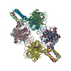 PDB-8s50: |