Biotechnology and Biological Sciences Research Council (BBSRC)
BB/T001542/1
United Kingdom
Biotechnology and Biological Sciences Research Council (BBSRC)
BB/T017643/1
United Kingdom
Citation
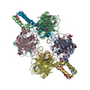
Journal: Matrix Biol / Year: 2025 Title: The structural organisation of pentraxin-3 and its interactions with heavy chains of inter-α-inhibitor regulate crosslinking of the hyaluronan matrix. Authors: Anokhi Shah / Xiaoli Zhang / Matthew Snee / Michael P Lockhart-Cairns / Colin W Levy / Thomas A Jowitt / Holly L Birchenough / Louisa Dean / Richard Collins / Rebecca J Dodd / Abigail R E ...Authors: Anokhi Shah / Xiaoli Zhang / Matthew Snee / Michael P Lockhart-Cairns / Colin W Levy / Thomas A Jowitt / Holly L Birchenough / Louisa Dean / Richard Collins / Rebecca J Dodd / Abigail R E Roberts / Jan J Enghild / Alberto Mantovani / Juan Fontana / Clair Baldock / Antonio Inforzato / Ralf P Richter / Anthony J Day / Abstract: Pentraxin-3 (PTX3) is an octameric protein, comprised of eight identical protomers, that has diverse functions in reproductive biology, innate immunity and cancer. PTX3 interacts with the large ...Pentraxin-3 (PTX3) is an octameric protein, comprised of eight identical protomers, that has diverse functions in reproductive biology, innate immunity and cancer. PTX3 interacts with the large polysaccharide hyaluronan (HA) to which heavy chains (HCs) of the inter-α-inhibitor (IαI) family of proteoglycans are covalently attached, playing a key role in the (non-covalent) crosslinking of HC•HA complexes. These interactions stabilise the cumulus matrix, essential for ovulation and fertilisation in mammals, and are also implicated in the formation of pathogenic matrices in the context of viral lung infections. To better understand the physiological and pathological roles of PTX3 we have analysed how its quaternary structure underpins HA crosslinking via its interactions with HCs. A combination of X-ray crystallography, cryo-electron microscopy (cryo-EM) and AlphaFold predictive modelling revealed that the C-terminal pentraxin domains of the PTX3 octamer are arranged in a central cube, with two long extensions on either side, each formed from four protomers assembled into tetrameric coiled-coil regions, essentially as described by (Noone et al., 2022; doi:10.1073/pnas.2208144119). From crystallography and cryo-EM data, we identified a network of inter-protomer salt bridges that facilitate the assembly of the octamer. Small angle X-ray scattering (SAXS) validated our model for the octameric protein, including the analysis of two PTX3 constructs: a tetrameric 'Half-PTX3' and a construct missing the 24 N-terminal residues (Δ1-24_PTX3). SAXS determined a length of ∼520 Å for PTX3 and, combined with 3D variability analysis of cryo-EM data, defined the flexibility of the N-terminal extensions. Biophysical analyses revealed that the prototypical heavy chain HC1 does not interact with PTX3 at pH 7.4, consistent with our previous studies showing that, at this pH, PTX3 only associates with HC•HA complexes if they are formed in its presence. However, PTX3 binds to HC1 at acidic pH, and can also be incorporated into pre-formed HC•HA complexes under these conditions. This provides a novel mechanism for the regulation of PTX3-mediated HA crosslinking (e.g., during inflammation), likely mediated by a pH-dependent conformational change in HC1. The PTX3 octamer was found to associate simultaneously with up to eight HC1 molecules and, thus, has the potential to form a major crosslinking node within HC•HA matrices, i.e., where the physical and biochemical properties of resulting matrices could be tuned by the HC/PTX3 composition.
Mass: 18.015 Da / Num. of mol.: 21 / Source method: isolated from a natural source / Formula: H2O
Has protein modification
Y
-
Experimental details
-
Experiment
Experiment
Method: X-RAY DIFFRACTION / Number of used crystals: 1
-
Sample preparation
Crystal
Density Matthews: 2.82 Å3/Da / Density % sol: 56.36 %
Crystal grow
Temperature: 277 K / Method: vapor diffusion, sitting drop Details: 10% w/v PEG 8000, 20% v/v ethylene glycol, 0.03 M of each halide, 0.1 M MOPS/HEPES-Na pH 7.5) * 0.3 M sodium fluoride, 0.3 M sodium bromide, 0.3 M sodium iodide Temp details: Cold room
-
Data collection
Diffraction
Mean temperature: 100 K / Serial crystal experiment: N
In the structure databanks used in Yorodumi, some data are registered as the other names, "COVID-19 virus" and "2019-nCoV". Here are the details of the virus and the list of structure data.
Jan 31, 2019. EMDB accession codes are about to change! (news from PDBe EMDB page)
EMDB accession codes are about to change! (news from PDBe EMDB page)
The allocation of 4 digits for EMDB accession codes will soon come to an end. Whilst these codes will remain in use, new EMDB accession codes will include an additional digit and will expand incrementally as the available range of codes is exhausted. The current 4-digit format prefixed with “EMD-” (i.e. EMD-XXXX) will advance to a 5-digit format (i.e. EMD-XXXXX), and so on. It is currently estimated that the 4-digit codes will be depleted around Spring 2019, at which point the 5-digit format will come into force.
The EM Navigator/Yorodumi systems omit the EMD- prefix.
Related info.:Q: What is EMD? / ID/Accession-code notation in Yorodumi/EM Navigator
Yorodumi is a browser for structure data from EMDB, PDB, SASBDB, etc.
This page is also the successor to EM Navigator detail page, and also detail information page/front-end page for Omokage search.
The word "yorodu" (or yorozu) is an old Japanese word meaning "ten thousand". "mi" (miru) is to see.
Related info.:EMDB / PDB / SASBDB / Comparison of 3 databanks / Yorodumi Search / Aug 31, 2016. New EM Navigator & Yorodumi / Yorodumi Papers / Jmol/JSmol / Function and homology information / Changes in new EM Navigator and Yorodumi
 Movie
Movie Controller


 Open data
 Basic information
 Components
 Keywords
 Function and homology information
 Homo sapiens (human)
 X-RAY DIFFRACTION /
  Authors
 United Kingdom, 2items
  Citation

 Structure visualization
 Downloads & links
 Other downloads
 PDBj
PDBj Assembly

 Cricetulus griseus (Chinese hamster) / References: UniProt: P26022
 Mass: 18.015 Da / Num. of mol.: 21 / Source method: isolated from a natural source / Formula: H2O
 Sample preparation
 Processing