 ムービー
ムービー コントローラー


+ データを開く

- 基本情報
基本情報
| 登録情報 | データベース: EMDB / ID: EMD-1943 | |||||||||
|---|---|---|---|---|---|---|---|---|---|---|
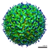
| タイトル | Feline Calicivirus strain F9 decorated with Junctional Adhesion Molecule A | |||||||||
 マップデータ マップデータ | Three-dimensional reconstruction of feline calicivirus bound to junctional adhesion molecule A prior to conformational changes | |||||||||
 試料 試料 |
| |||||||||
 キーワード キーワード | feline / calicivirus / virus / virion / junctional adhesion molecule | |||||||||
| 生物種 |   Feline calicivirus (ウイルス) Feline calicivirus (ウイルス) | |||||||||
| 手法 | 単粒子再構成法 / クライオ電子顕微鏡法 / 解像度: 11.8 Å | |||||||||
 データ登録者 データ登録者 | Bhella D / Goodfellow IG | |||||||||
 引用 引用 | ジャーナル: J Virol / 年: 2011 タイトル: The cryo-electron microscopy structure of feline calicivirus bound to junctional adhesion molecule A at 9-angstrom resolution reveals receptor-induced flexibility and two distinct ...タイトル: The cryo-electron microscopy structure of feline calicivirus bound to junctional adhesion molecule A at 9-angstrom resolution reveals receptor-induced flexibility and two distinct conformational changes in the capsid protein VP1. 著者: David Bhella / Ian G Goodfellow /  要旨: Caliciviridae are small icosahedral positive-sense RNA-containing viruses and include the human noroviruses, a leading cause of infectious acute gastroenteritis and feline calicivirus (FCV), which ...Caliciviridae are small icosahedral positive-sense RNA-containing viruses and include the human noroviruses, a leading cause of infectious acute gastroenteritis and feline calicivirus (FCV), which causes respiratory illness and stomatitis in cats. FCV attachment and entry is mediated by feline junctional adhesion molecule A (fJAM-A), which binds to the outer face of the capsomere, inducing a conformational change in the capsid that may be important for viral uncoating. Here we present the results of our structural investigation of the virus-receptor interaction and ensuing conformational changes. Cryo-electron microscopy and three-dimensional image reconstruction were used to solve the structure of the virus decorated with a soluble fragment of the receptor at subnanometer resolution. In initial reconstructions, the P domains of the capsid protein VP1 and fJAM-A were poorly resolved. Sorting experiments led to improved reconstructions of the FCV-fJAM-A complex both before and after the induced conformational change, as well as in three transition states. These data showed that the P domain becomes flexible following fJAM-A binding, leading to a loss of icosahedral symmetry. Furthermore, two distinct conformational changes were seen; an anticlockwise rotation of up to 15° of the P domain was observed in the AB dimers, while tilting of the P domain away from the icosahedral 2-fold axis was seen in the CC dimers. A list of putative contact residues was calculated by fitting high-resolution coordinates for fJAM-A and VP1 to the reconstructed density maps, highlighting regions in both virus and receptor important for virus attachment and entry. | |||||||||
| 履歴 |
|
- 構造の表示
構造の表示
| ムービー |
 ムービービューア ムービービューア |
|---|---|
| 構造ビューア | EMマップ: SurfViewMolmilJmol/JSmol |
| 添付画像 |

- ダウンロードとリンク
-EMDBアーカイブ
| マップデータ | emd_1943.map.gz | 15.2 MB | EMDBマップデータ形式 | |
|---|---|---|---|---|
| ヘッダ (付随情報) | emd-1943-v30.xmlemd-1943.xml | 10.1 KB 10.1 KB | 表示 表示 | EMDBヘッダ |
| 画像 | 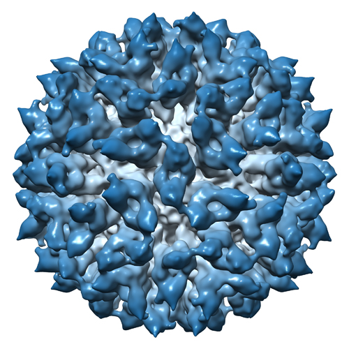 EMD-1943.jpg EMD-1943.jpg | 225.5 KB | ||
| アーカイブディレクトリ |  http://ftp.pdbj.org/pub/emdb/structures/EMD-1943ftp://ftp.pdbj.org/pub/emdb/structures/EMD-1943 http://ftp.pdbj.org/pub/emdb/structures/EMD-1943ftp://ftp.pdbj.org/pub/emdb/structures/EMD-1943 | HTTPS FTP |
-検証レポート
| 文書・要旨 | emd_1943_validation.pdf.gz | 261.5 KB | 表示 | EMDB検証レポート |
|---|---|---|---|---|
| 文書・詳細版 | emd_1943_full_validation.pdf.gz | 260.7 KB | 表示 | |
| XML形式データ | emd_1943_validation.xml.gz | 12.7 KB | 表示 | |
| アーカイブディレクトリ | https://ftp.pdbj.org/pub/emdb/validation_reports/EMD-1943ftp://ftp.pdbj.org/pub/emdb/validation_reports/EMD-1943 | HTTPS FTP |
-関連構造データ


-リンク
| EMDBのページ | EMDB (EBI/PDBe) / EMDataResource |
|---|
-マップ
| ファイル | ダウンロード / ファイル: emd_1943.map.gz / 形式: CCP4 / 大きさ: 49.8 MB / タイプ: IMAGE STORED AS SIGNED INTEGER (2 BYTES) | ||||||||||||||||||||||||||||||||||||||||||||||||||||||||||||||||||||
|---|---|---|---|---|---|---|---|---|---|---|---|---|---|---|---|---|---|---|---|---|---|---|---|---|---|---|---|---|---|---|---|---|---|---|---|---|---|---|---|---|---|---|---|---|---|---|---|---|---|---|---|---|---|---|---|---|---|---|---|---|---|---|---|---|---|---|---|---|---|
| 注釈 | Three-dimensional reconstruction of feline calicivirus bound to junctional adhesion molecule A prior to conformational changes | ||||||||||||||||||||||||||||||||||||||||||||||||||||||||||||||||||||
| 投影像・断面図 | 画像のコントロール
画像は Spider により作成 | ||||||||||||||||||||||||||||||||||||||||||||||||||||||||||||||||||||
| ボクセルのサイズ | X=Y=Z: 2.06 Å | ||||||||||||||||||||||||||||||||||||||||||||||||||||||||||||||||||||
| 密度 |
| ||||||||||||||||||||||||||||||||||||||||||||||||||||||||||||||||||||
| 対称性 | 空間群: 1 | ||||||||||||||||||||||||||||||||||||||||||||||||||||||||||||||||||||
| 詳細 | EMDB XML:
CCP4マップ ヘッダ情報:
| ||||||||||||||||||||||||||||||||||||||||||||||||||||||||||||||||||||
 Z (Sec.)
 Y (Row.)
 X (Col.)


-添付データ
- 試料の構成要素
-全体 : Feline Calicivirus decorated with junctional adhesion molecule A
| 全体 | 名称: Feline Calicivirus decorated with junctional adhesion molecule A |
|---|---|
| 要素 |
|
-超分子 #1000: Feline Calicivirus decorated with junctional adhesion molecule A
| 超分子 | 名称: Feline Calicivirus decorated with junctional adhesion molecule A タイプ: sample / ID: 1000 / 集合状態: T3 icosahedral capsid / Number unique components: 2 |
|---|---|
| 分子量 | 理論値: 16 MDa |
-超分子 #1: Feline calicivirus
| 超分子 | 名称: Feline calicivirus / タイプ: virus / ID: 1 / Name.synonym: Feline calicivirus / NCBI-ID: 11978 / 生物種: Feline calicivirus / ウイルスタイプ: VIRION / ウイルス・単離状態: STRAIN / ウイルス・エンベロープ: No / ウイルス・中空状態: No / Syn species name: Feline calicivirus |
|---|---|
| 宿主 | 生物種: |
| 分子量 | 理論値: 10.7 MDa |
| ウイルス殻 | Shell ID: 1 / 名称: VP1 / 直径: 415 Å / T番号(三角分割数): 3 |
-分子 #1: Junctional Adhesion Molecule A
| 分子 | 名称: Junctional Adhesion Molecule A / タイプ: protein_or_peptide / ID: 1 / Name.synonym: fJAM-A 詳細: Virus was incubated in the presence of a soluble fragment of fJAM-A at 4oC for one hour コピー数: 180 / 集合状態: Monomer / 組換発現: Yes |
|---|---|
| 由来(天然) | 生物種: |
| 分子量 | 理論値: 29 KDa |
| 組換発現 | 生物種:  Cricetulus griseus (モンゴルキヌゲネズミ) Cricetulus griseus (モンゴルキヌゲネズミ)組換プラスミド: pDEF:fJAM:Fc |
-実験情報
-構造解析
| 手法 | クライオ電子顕微鏡法 |
|---|---|
 解析 解析 | 単粒子再構成法 |
| 試料の集合状態 | particle |
-試料調製
| 緩衝液 | pH: 7.4 / 詳細: PBS |
|---|---|
| グリッド | 詳細: 400 mesh R2/2 C-flat |
| 凍結 | 凍結剤: ETHANE / チャンバー内湿度: 100 % / 装置: OTHER / 詳細: Vitrification instrument: Vitrobot / 手法: blot for five seconds before plunging |
- 電子顕微鏡法
| 顕微鏡 | JEOL 2200FS |
|---|---|
| 温度 | 平均: 97 K |
| アライメント法 | Legacy - 非点収差: corrected at 100k times mag in digital micrograph |
| 特殊光学系 | エネルギーフィルター - 名称: JEOL エネルギーフィルター - エネルギー下限: 0.0 eV エネルギーフィルター - エネルギー上限: 20.0 eV |
| 撮影 | カテゴリ: CCD フィルム・検出器のモデル: GATAN ULTRASCAN 4000 (4k x 4k) 平均電子線量: 10 e/Å2 |
| 電子線 | 加速電圧: 200 kV / 電子線源:  FIELD EMISSION GUN FIELD EMISSION GUN |
| 電子光学系 | 照射モード: FLOOD BEAM / 撮影モード: BRIGHT FIELD / Cs: 2.0 mm / 最大 デフォーカス(公称値): 3.5 µm / 最小 デフォーカス(公称値): 0.8 µm / 倍率(公称値): 100000 |
| 試料ステージ | 試料ホルダー: Side entry / 試料ホルダーモデル: GATAN LIQUID NITROGEN |
-画像解析
| CTF補正 | 詳細: each particle corrected using bsoft |
|---|---|
| 最終 再構成 | 想定した対称性 - 点群: I (正20面体型対称) / アルゴリズム: OTHER / 解像度のタイプ: BY AUTHOR / 解像度: 11.8 Å / 解像度の算出法: FSC 0.5 CUT-OFF / ソフトウェア - 名称: em3dr2 / 使用した粒子像数: 862 |
