 Movie
Movie Controller


+ Open data

- Basic information
| Entry | Database: EMDB / ID: EMD-1941 | |||||||||
|---|---|---|---|---|---|---|---|---|---|---|
| Title | Radial Spoke from Chlamydomonas flagella | |||||||||
 Map data Map data | This is a density map of Radial Spokes from Chlamydomonas flagella. | |||||||||
 Sample Sample |
| |||||||||
 Keywords Keywords | radial spoke / flagella / cilia / Chlamydomonas | |||||||||
| Biological species |   Chlamydomonas reinhardtii (plant) Chlamydomonas reinhardtii (plant) | |||||||||
| Method | subtomogram averaging / cryo EM / Resolution: 39.18 Å | |||||||||
 Authors Authors | Pigino G / Bui KH / Maheshwari A / Lupetti P / Diener D / Ishikawa T | |||||||||
 Citation Citation | Journal: J Cell Biol / Year: 2011 Title: Cryoelectron tomography of radial spokes in cilia and flagella. Authors: Gaia Pigino / Khanh Huy Bui / Aditi Maheshwari / Pietro Lupetti / Dennis Diener / Takashi Ishikawa /  Abstract: Radial spokes (RSs) are ubiquitous components in the 9 + 2 axoneme thought to be mechanochemical transducers involved in local control of dynein-driven microtubule sliding. They are composed of >23 ...Radial spokes (RSs) are ubiquitous components in the 9 + 2 axoneme thought to be mechanochemical transducers involved in local control of dynein-driven microtubule sliding. They are composed of >23 polypeptides, whose interactions and placement must be deciphered to understand RS function. In this paper, we show the detailed three-dimensional (3D) structure of RS in situ in Chlamydomonas reinhardtii flagella and Tetrahymena thermophila cilia that we obtained using cryoelectron tomography (cryo-ET). We clarify similarities and differences between the three spoke species, RS1, RS2, and RS3, in T. thermophila and in C. reinhardtii and show that part of RS3 is conserved in C. reinhardtii, which only has two species of complete RSs. By analyzing C. reinhardtii mutants, we identified the specific location of subsets of RS proteins (RSPs). Our 3D reconstructions show a twofold symmetry, suggesting that fully assembled RSs are produced by dimerization. Based on our cryo-ET data, we propose models of subdomain organization within the RS as well as interactions between RSPs and with other axonemal components. | |||||||||
| History |
|
- Structure visualization
| Movie |
 Movie viewer Movie viewer |
|---|---|
| Structure viewer | EM map: SurfViewMolmilJmol/JSmol |
| Supplemental images |

- Downloads & links
-EMDB archive
| Map data | emd_1941.map.gz | 787.5 KB | EMDB map data format | |
|---|---|---|---|---|
| Header (meta data) | emd-1941-v30.xmlemd-1941.xml | 9.6 KB 9.6 KB | Display Display | EMDB header |
| Images | 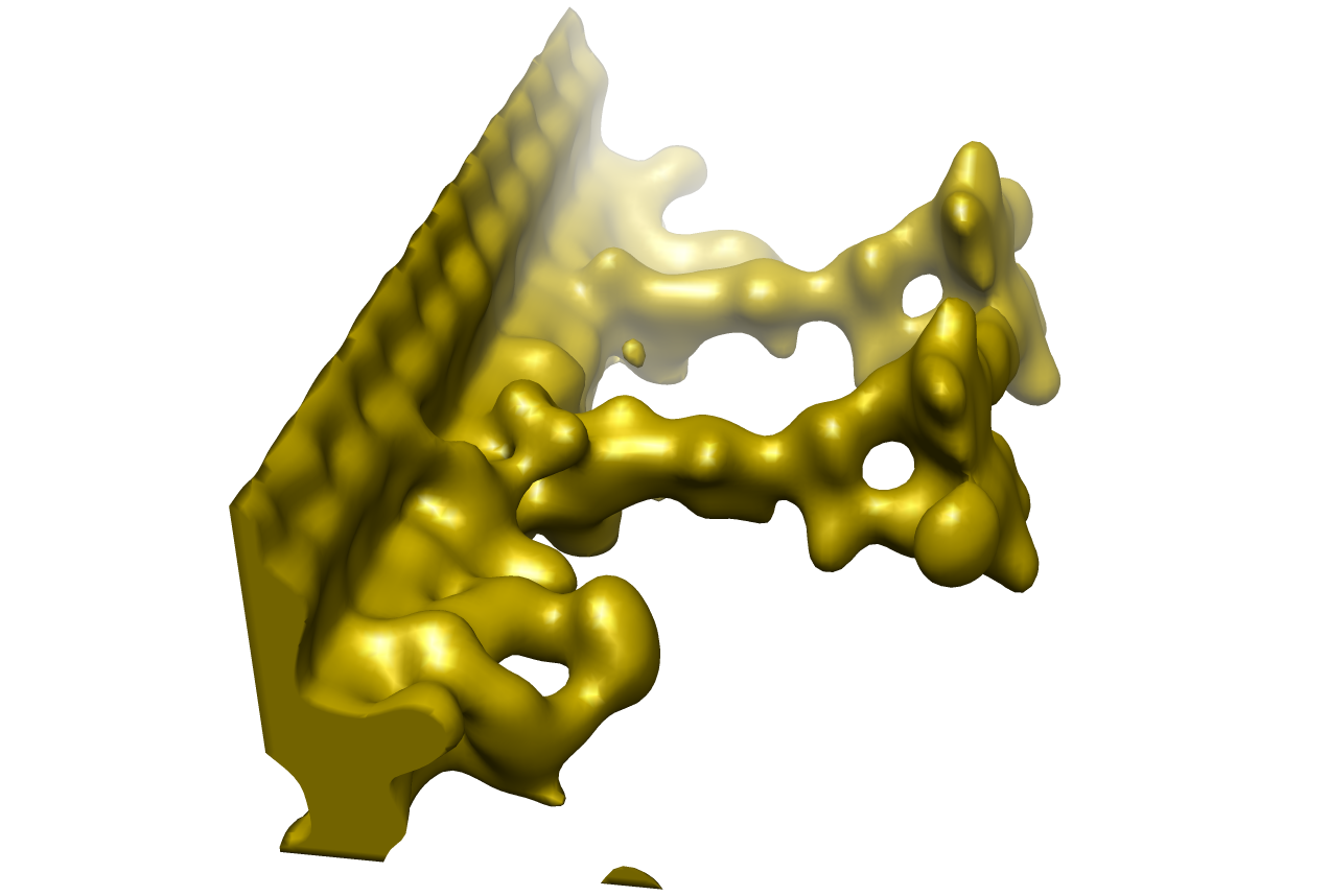 1941.png 1941.png | 270.2 KB | ||
| Archive directory |  http://ftp.pdbj.org/pub/emdb/structures/EMD-1941ftp://ftp.pdbj.org/pub/emdb/structures/EMD-1941 http://ftp.pdbj.org/pub/emdb/structures/EMD-1941ftp://ftp.pdbj.org/pub/emdb/structures/EMD-1941 | HTTPS FTP |
-Validation report
| Summary document | emd_1941_validation.pdf.gz | 175.4 KB | Display | EMDB validaton report |
|---|---|---|---|---|
| Full document | emd_1941_full_validation.pdf.gz | 174.5 KB | Display | |
| Data in XML | emd_1941_validation.xml.gz | 4.8 KB | Display | |
| Arichive directory | https://ftp.pdbj.org/pub/emdb/validation_reports/EMD-1941ftp://ftp.pdbj.org/pub/emdb/validation_reports/EMD-1941 | HTTPS FTP |
-Related structure data
| Similar structure data |
|---|
-Links
| EMDB pages | EMDB (EBI/PDBe) / EMDataResource |
|---|
-Map
| File | Download / File: emd_1941.map.gz / Format: CCP4 / Size: 2 MB / Type: IMAGE STORED AS FLOATING POINT NUMBER (4 BYTES) | ||||||||||||||||||||||||||||||||||||||||||||||||||||||||||||||||||||
|---|---|---|---|---|---|---|---|---|---|---|---|---|---|---|---|---|---|---|---|---|---|---|---|---|---|---|---|---|---|---|---|---|---|---|---|---|---|---|---|---|---|---|---|---|---|---|---|---|---|---|---|---|---|---|---|---|---|---|---|---|---|---|---|---|---|---|---|---|---|
| Annotation | This is a density map of Radial Spokes from Chlamydomonas flagella. | ||||||||||||||||||||||||||||||||||||||||||||||||||||||||||||||||||||
| Projections & slices | Image control
Images are generated by Spider. generated in cubic-lattice coordinate | ||||||||||||||||||||||||||||||||||||||||||||||||||||||||||||||||||||
| Voxel size | X=Y=Z: 6.78 Å | ||||||||||||||||||||||||||||||||||||||||||||||||||||||||||||||||||||
| Density |
| ||||||||||||||||||||||||||||||||||||||||||||||||||||||||||||||||||||
| Symmetry | Space group: 1 | ||||||||||||||||||||||||||||||||||||||||||||||||||||||||||||||||||||
| Details | EMDB XML:
CCP4 map header:
| ||||||||||||||||||||||||||||||||||||||||||||||||||||||||||||||||||||
 Z (Sec.)
 Y (Row.)
 X (Col.)


-Supplemental data
- Sample components
-Entire : Radial spoke from flagella/cilia
| Entire | Name: Radial spoke from flagella/cilia |
|---|---|
| Components |
|
-Supramolecule #1000: Radial spoke from flagella/cilia
| Supramolecule | Name: Radial spoke from flagella/cilia / type: sample / ID: 1000 / Details: Radial spokes from Axoneme / Number unique components: 1 |
|---|
-Supramolecule #1: radial spoke
| Supramolecule | Name: radial spoke / type: organelle_or_cellular_component / ID: 1 / Name.synonym: radial spoke / Recombinant expression: Yes |
|---|---|
| Source (natural) | Organism: Chlamydomonas reinhardtii (plant) / Strain: 137c / synonym: Chlamydomonas / Tissue: flagella / Cell: 137c / Organelle: flagella / Location in cell: flagella |
| Recombinant expression | Organism: Chlamydomonas reinhardtii (plant) |
-Experimental details
-Structure determination
| Method | cryo EM |
|---|---|
 Processing Processing | subtomogram averaging |
-Sample preparation
| Concentration | 1 mg/mL |
|---|---|
| Buffer | pH: 7.4 / Details: HMDKP |
| Grid | Details: lacey carbon grids |
| Vitrification | Cryogen name: ETHANE / Chamber humidity: 90 % / Chamber temperature: 100 K / Instrument: OTHER / Details: Vitrification instrument: Vitrobot / Method: 3 second blotting |
- Electron microscopy
| Microscope | FEI TECNAI 20 |
|---|---|
| Temperature | Min: 100 K / Max: 100 K / Average: 100 K |
| Specialist optics | Energy filter - Name: GIF / Energy filter - Lower energy threshold: 0.0 eV / Energy filter - Upper energy threshold: 20.0 eV |
| Image recording | Category: CCD / Film or detector model: GATAN ULTRASCAN 1000 (2k x 2k) / Average electron dose: 0.6 e/Å2 |
| Electron beam | Acceleration voltage: 200 kV / Electron source:  FIELD EMISSION GUN FIELD EMISSION GUN |
| Electron optics | Calibrated magnification: 19000 / Illumination mode: FLOOD BEAM / Imaging mode: BRIGHT FIELD / Cs: 1.5 mm / Nominal defocus max: 6.0 µm / Nominal defocus min: 3.0 µm / Nominal magnification: 27500 |
| Sample stage | Specimen holder: Gatan 626 / Specimen holder model: GATAN LIQUID NITROGEN / Tilt series - Axis1 - Min angle: -60 ° / Tilt series - Axis1 - Max angle: 60 ° |
-Image processing
| Details | Subtomograms were averaged. Average number of tilts used in the 3D reconstructions: 61. Average tomographic tilt angle increment: 2. |
|---|---|
| Final reconstruction | Algorithm: OTHER / Resolution.type: BY AUTHOR / Resolution: 39.18 Å / Resolution method: FSC 0.5 CUT-OFF / Software - Name: IMOD |
