Movie
Movie Controller


+ Open data

- Basic information
| Entry | Database: EMDB / ID: EMD-1931 | |||||||||
|---|---|---|---|---|---|---|---|---|---|---|
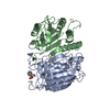
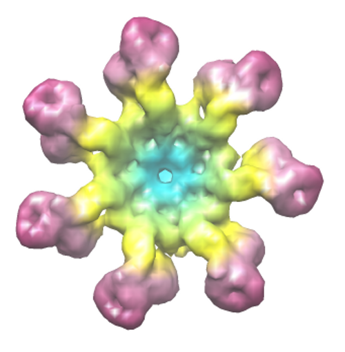
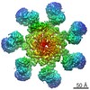
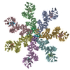
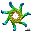
| Title | Map of the apoptosome-procaspase-9 complex | |||||||||
 Map data Map data | Map of the apoptosome-procaspase-9 complex | |||||||||
 Sample Sample |
| |||||||||
 Keywords Keywords | apoptosome / Apaf-1 / procaspase-9 activation | |||||||||
| Function / homology | Apoptotic protease-activating factor 1 Function and homology information Function and homology information | |||||||||
| Biological species |  Homo sapiens (human) / Homo sapiens (human) /  | |||||||||
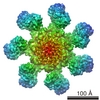
| Method | single particle reconstruction / cryo EM / Resolution: 16.9 Å | |||||||||
 Authors Authors | Yuan S / Ludtke SJ / Akey CW | |||||||||
 Citation Citation | Journal: Structure / Year: 2011 Title: The holo-apoptosome: activation of procaspase-9 and interactions with caspase-3. Authors: Shujun Yuan / Xinchao Yu / John M Asara / John E Heuser / Steven J Ludtke / Christopher W Akey /  Abstract: Activation of procaspase-9 on the apoptosome is a pivotal step in the intrinsic cell death pathway. We now provide further evidence that caspase recruitment domains of pc-9 and Apaf-1 form a CARD- ...Activation of procaspase-9 on the apoptosome is a pivotal step in the intrinsic cell death pathway. We now provide further evidence that caspase recruitment domains of pc-9 and Apaf-1 form a CARD-CARD disk that is flexibly tethered to the apoptosome. In addition, a 3D reconstruction of the pc-9 apoptosome was calculated without symmetry restraints. In this structure, p20 and p10 catalytic domains of a single pc-9 interact with nucleotide binding domains of adjacent Apaf-1 subunits. Together, disk assembly and pc-9 binding create an asymmetric proteolysis machine. We also show that CARD-p20 and p20-p10 linkers play important roles in pc-9 activation. Based on the data, we propose a proximity-induced association model for pc-9 activation on the apoptosome. We also show that pc-9 and caspase-3 have overlapping binding sites on the central hub. These binding sites may play a role in pc-3 activation and could allow the formation of hybrid apoptosomes with pc-9 and caspase-3 proteolytic activities. | |||||||||
| History |
|
- Structure visualization
| Movie |
Movie viewer |
|---|---|
| Structure viewer | EM map: SurfViewMolmilJmol/JSmol |
| Supplemental images |
- Downloads & links
-EMDB archive
| Map data | emd_1931.map.gz | 24.5 MB | EMDB map data format | |
|---|---|---|---|---|
| Header (meta data) | emd-1931-v30.xmlemd-1931.xml | 12.7 KB 12.7 KB | Display Display | EMDB header |
| Images |  emd_1931.png emd_1931.png | 157.6 KB | ||
| Archive directory |  http://ftp.pdbj.org/pub/emdb/structures/EMD-1931ftp://ftp.pdbj.org/pub/emdb/structures/EMD-1931 http://ftp.pdbj.org/pub/emdb/structures/EMD-1931ftp://ftp.pdbj.org/pub/emdb/structures/EMD-1931 | HTTPS FTP |
-Related structure data
| Similar structure data |
|---|

-Links
| EMDB pages | EMDB (EBI/PDBe) / EMDataResource |
|---|
-Map
| File | Download / File: emd_1931.map.gz / Format: CCP4 / Size: 26.4 MB / Type: IMAGE STORED AS FLOATING POINT NUMBER (4 BYTES) | ||||||||||||||||||||||||||||||||||||||||||||||||||||||||||||||||||||
|---|---|---|---|---|---|---|---|---|---|---|---|---|---|---|---|---|---|---|---|---|---|---|---|---|---|---|---|---|---|---|---|---|---|---|---|---|---|---|---|---|---|---|---|---|---|---|---|---|---|---|---|---|---|---|---|---|---|---|---|---|---|---|---|---|---|---|---|---|---|
| Annotation | Map of the apoptosome-procaspase-9 complex | ||||||||||||||||||||||||||||||||||||||||||||||||||||||||||||||||||||
| Projections & slices | Image control
Images are generated by Spider. | ||||||||||||||||||||||||||||||||||||||||||||||||||||||||||||||||||||
| Voxel size | X=Y=Z: 2.9 Å | ||||||||||||||||||||||||||||||||||||||||||||||||||||||||||||||||||||
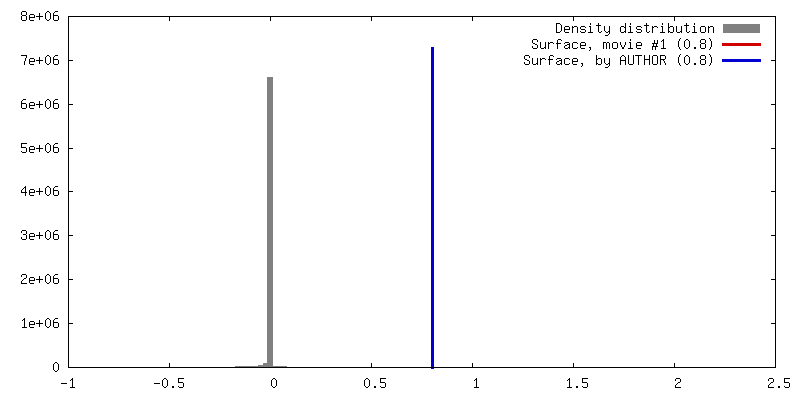
| Density |
| ||||||||||||||||||||||||||||||||||||||||||||||||||||||||||||||||||||
| Symmetry | Space group: 1 | ||||||||||||||||||||||||||||||||||||||||||||||||||||||||||||||||||||
| Details | EMDB XML:
CCP4 map header:
| ||||||||||||||||||||||||||||||||||||||||||||||||||||||||||||||||||||
 Z (Sec.)
 Y (Row.)
 X (Col.)


-Supplemental data
- Sample components
-Entire : Human apoptosome-procaspase-9 complex
| Entire | Name: Human apoptosome-procaspase-9 complex |
|---|---|
| Components |
|
-Supramolecule #1000: Human apoptosome-procaspase-9 complex
| Supramolecule | Name: Human apoptosome-procaspase-9 complex / type: sample / ID: 1000 Details: A slight excess of procaspase-9 was added to ensure saturation of binding to sites on the apoptosome. Oligomeric state: 5-7 procaspase-9 molecules bound to the human apoptosome comprised of 7 Apaf-1 subunits Number unique components: 3 |
|---|---|
| Molecular weight | Theoretical: 1.2 MDa |
-Macromolecule #1: Apaf-1
| Macromolecule | Name: Apaf-1 / type: protein_or_peptide / ID: 1 / Name.synonym: Apaf-1 Details: Apaf-1, cytochrome c and procaspase-9 were co-assembled in the presence of dATP to form the apoptosome-procaspase-9 complex Number of copies: 7 / Oligomeric state: Heptamer / Recombinant expression: Yes |
|---|---|
| Source (natural) | Organism: Homo sapiens (human) / synonym: Human / Location in cell: Cytosol |
| Molecular weight | Theoretical: 130 KDa |
| Recombinant expression | Organism:   Spodoptera frugiperda (fall armyworm) / Recombinant plasmid: pFastBac1 Spodoptera frugiperda (fall armyworm) / Recombinant plasmid: pFastBac1 |
| Sequence | InterPro: Apoptotic protease-activating factor 1 |
-Macromolecule #2: Procaspase-9
| Macromolecule | Name: Procaspase-9 / type: protein_or_peptide / ID: 2 / Name.synonym: Procaspase-9 / Oligomeric state: Monomer / Recombinant expression: Yes |
|---|---|
| Source (natural) | Organism: Homo sapiens (human) / synonym: Human / Location in cell: Cytosol |
| Molecular weight | Theoretical: 50 KDa |
| Recombinant expression | Organism:  |
-Macromolecule #3: Cytochrome c
| Macromolecule | Name: Cytochrome c / type: protein_or_peptide / ID: 3 / Name.synonym: Cytochrome c / Number of copies: 7 / Oligomeric state: Monomer / Recombinant expression: No |
|---|---|
| Source (natural) | Organism: |
| Molecular weight | Theoretical: 10 KDa |
-Experimental details
-Structure determination
| Method | cryo EM |
|---|---|
 Processing Processing | single particle reconstruction |
| Aggregation state | particle |
-Sample preparation
| Concentration | 3 mg/mL |
|---|---|
| Buffer | pH: 7.5 Details: 20 mM HEPES, 10 mM KCl, 1.5 mM MgCl2, 1 mM EDTA, 1 mM EGTA, 1 mM DTT |
| Grid | Details: Quantifoil holey grids |
| Vitrification | Cryogen name: ETHANE / Chamber humidity: 100 % / Chamber temperature: 77 K / Instrument: FEI VITROBOT MARK III / Details: Vitrification instrument: Vitrobot Mark 3 (FEI) / Method: Blot for 2-3 seconds before plunging at 20C |
- Electron microscopy
| Microscope | FEI TECNAI F20 |
|---|---|
| Temperature | Average: 96 K |
| Alignment procedure | Legacy - Astigmatism: objective lens astigmatism was corrected at 200,000 times magnification |
| Details | 4k x 4k ccd used |
| Image recording | Category: CCD / Film or detector model: GENERIC TVIPS / Number real images: 447 / Average electron dose: 20 e/Å2 |
| Electron beam | Acceleration voltage: 120 kV / Electron source:  FIELD EMISSION GUN FIELD EMISSION GUN |
| Electron optics | Calibrated magnification: 29000 / Illumination mode: FLOOD BEAM / Imaging mode: BRIGHT FIELD / Cs: 2 mm / Nominal defocus max: 3.5 µm / Nominal defocus min: 1.8 µm / Nominal magnification: 29000 |
| Sample stage | Specimen holder: Side entry liquid nitrogen-cooled cryo specimen holder Specimen holder model: GATAN LIQUID NITROGEN |
| Experimental equipment |  Model: Tecnai F20 / Image courtesy: FEI Company |
-Image processing
| Details | Apaf-1 was co-assembled with bovine cytochrome c and pc-9 |
|---|---|
| CTF correction | Details: Each image |
| Final reconstruction | Applied symmetry - Point group: C1 (asymmetric) / Algorithm: OTHER / Resolution.type: BY AUTHOR / Resolution: 16.9 Å / Resolution method: FSC 0.5 CUT-OFF / Software - Name: EMAN2 Details: 3D volume was calculated without imposing any symmetry (c1) Number images used: 20000 |
| Final two d classification | Number classes: 1000 |
-Atomic model buiding 1
| Initial model | PDB ID:  3iza |
|---|---|
| Software | Name: chimera |
| Details | Protocol: rigid body |
| Refinement | Space: REAL / Protocol: RIGID BODY FIT / Target criteria: cross correlation |
-Atomic model buiding 2
| Initial model | PDB ID: Chain - Chain ID: A |
|---|---|
| Software | Name: chimera |
| Details | PDBEntryID_givenInChain. Protocol: rigid body. final fit was done manually taking into account the orientation of the p20-p10 loop. |
| Refinement | Space: REAL / Protocol: RIGID BODY FIT |