 ムービー
ムービー コントローラー


+ データを開く

- 基本情報
基本情報
| 登録情報 | データベース: EMDB / ID: EMD-1907 | |||||||||
|---|---|---|---|---|---|---|---|---|---|---|
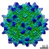
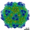
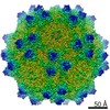
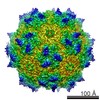
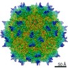
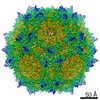
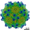
| タイトル | Electron cryo-microscopy and image reconstruction of adeno-associated virus type 2 empty capsids | |||||||||
 マップデータ マップデータ | AAV-2 empty capsids | |||||||||
 試料 試料 |
| |||||||||
 キーワード キーワード | AAV2 / virus / empty capsids | |||||||||
| 生物種 |  Adeno-associated virus - 2 (アデノ随伴ウイルス) Adeno-associated virus - 2 (アデノ随伴ウイルス) | |||||||||
| 手法 | 単粒子再構成法 / クライオ電子顕微鏡法 / 解像度: 10.5 Å | |||||||||
 データ登録者 データ登録者 | Kronenberg S / Kleinschmidt JA / Bottcher B | |||||||||
 引用 引用 | ジャーナル: EMBO Rep / 年: 2001 タイトル: Electron cryo-microscopy and image reconstruction of adeno-associated virus type 2 empty capsids. 著者: S Kronenberg / J A Kleinschmidt / B Böttcher /  要旨: Adeno-associated virus type 2 empty capsids are composed of three proteins, VP1, VP2 and VP3, which have relative molecular masses of 87, 72 and 62 kDa, respectively, and differ in their N-terminal ...Adeno-associated virus type 2 empty capsids are composed of three proteins, VP1, VP2 and VP3, which have relative molecular masses of 87, 72 and 62 kDa, respectively, and differ in their N-terminal amino acid sequences. They have a likely molar ratio of 1:1:8 and occupy symmetrical equivalent positions in an icosahedrally arranged protein shell. We have investigated empty capsids of adeno-associated virus type 2 by electron cryo-microscopy and icosahedral image reconstruction. The three-dimensional map at 1.05 nm resolution showed sets of three elongated spikes surrounding the three-fold symmetry axes and narrow empty channels at the five-fold axes. The inside of the capsid superimposed with the previously determined structure of the canine parvovirus (Q. Xie and M.S. Chapman, 1996, J. Mol. Biol., 264, 497-520), whereas the outer surface showed clear discrepancies. Globular structures at the inner surface of the capsid at the two-fold symmetry axes were identified as possible positions for the N-terminal extensions of VP1 and VP2. | |||||||||
| 履歴 |
|
- 構造の表示
構造の表示
| ムービー |
 ムービービューア ムービービューア |
|---|---|
| 構造ビューア | EMマップ: SurfViewMolmilJmol/JSmol |
| 添付画像 |
 UCSF Chimera
- ダウンロードとリンク
-EMDBアーカイブ
| マップデータ | emd_1907.map.gz | 2 MB | EMDBマップデータ形式 | |
|---|---|---|---|---|
| ヘッダ (付随情報) | emd-1907-v30.xmlemd-1907.xml | 9.1 KB 9.1 KB | 表示 表示 | EMDBヘッダ |
| 画像 | 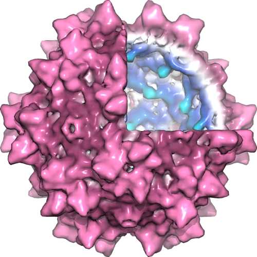 1907_aav2_1.jpg 1907_aav2_1.jpg | 26.9 KB | ||
| アーカイブディレクトリ |  http://ftp.pdbj.org/pub/emdb/structures/EMD-1907ftp://ftp.pdbj.org/pub/emdb/structures/EMD-1907 http://ftp.pdbj.org/pub/emdb/structures/EMD-1907ftp://ftp.pdbj.org/pub/emdb/structures/EMD-1907 | HTTPS FTP |
-検証レポート
| 文書・要旨 | emd_1907_validation.pdf.gz | 268.5 KB | 表示 | EMDB検証レポート |
|---|---|---|---|---|
| 文書・詳細版 | emd_1907_full_validation.pdf.gz | 267.7 KB | 表示 | |
| XML形式データ | emd_1907_validation.xml.gz | 5.6 KB | 表示 | |
| アーカイブディレクトリ | https://ftp.pdbj.org/pub/emdb/validation_reports/EMD-1907ftp://ftp.pdbj.org/pub/emdb/validation_reports/EMD-1907 | HTTPS FTP |
-関連構造データ
-リンク
| EMDBのページ | EMDB (EBI/PDBe) / EMDataResource |
|---|
-マップ
| ファイル | ダウンロード / ファイル: emd_1907.map.gz / 形式: CCP4 / 大きさ: 2.1 MB / タイプ: IMAGE STORED AS FLOATING POINT NUMBER (4 BYTES) | ||||||||||||||||||||||||||||||||||||||||||||||||||||||||||||||||||||
|---|---|---|---|---|---|---|---|---|---|---|---|---|---|---|---|---|---|---|---|---|---|---|---|---|---|---|---|---|---|---|---|---|---|---|---|---|---|---|---|---|---|---|---|---|---|---|---|---|---|---|---|---|---|---|---|---|---|---|---|---|---|---|---|---|---|---|---|---|---|
| 注釈 | AAV-2 empty capsids | ||||||||||||||||||||||||||||||||||||||||||||||||||||||||||||||||||||
| 投影像・断面図 | 画像のコントロール
画像は Spider により作成 | ||||||||||||||||||||||||||||||||||||||||||||||||||||||||||||||||||||
| ボクセルのサイズ | X=Y=Z: 4.2 Å | ||||||||||||||||||||||||||||||||||||||||||||||||||||||||||||||||||||
| 密度 |
| ||||||||||||||||||||||||||||||||||||||||||||||||||||||||||||||||||||
| 対称性 | 空間群: 1 | ||||||||||||||||||||||||||||||||||||||||||||||||||||||||||||||||||||
| 詳細 | EMDB XML:
CCP4マップ ヘッダ情報:
| ||||||||||||||||||||||||||||||||||||||||||||||||||||||||||||||||||||
 Z (Sec.)
 Y (Row.)
 X (Col.)


-添付データ
- 試料の構成要素
-全体 : Adeno-associated Virus Type 2
| 全体 | 名称: Adeno-associated Virus Type 2 |
|---|---|
| 要素 |
|
-超分子 #1000: Adeno-associated Virus Type 2
| 超分子 | 名称: Adeno-associated Virus Type 2 / タイプ: sample / ID: 1000 / Number unique components: 1 |
|---|---|
| 分子量 | 理論値: 3.9 MDa |
-超分子 #1: Adeno-associated virus - 2
| 超分子 | 名称: Adeno-associated virus - 2 / タイプ: virus / ID: 1 / Name.synonym: AAV2 / NCBI-ID: 10804 / 生物種: Adeno-associated virus - 2 / ウイルスタイプ: VIRUS-LIKE PARTICLE / ウイルス・単離状態: SEROTYPE / ウイルス・エンベロープ: No / ウイルス・中空状態: Yes / Syn species name: AAV2 |
|---|---|
| 宿主 | 生物種:  Homo sapiens (ヒト) / 別称: VERTEBRATES Homo sapiens (ヒト) / 別称: VERTEBRATES |
| 分子量 | 理論値: 3.9 MDa |
| ウイルス殻 | Shell ID: 1 / 名称: AAV2 / 直径: 260 Å / T番号(三角分割数): 1 |
-実験情報
-構造解析
| 手法 | クライオ電子顕微鏡法 |
|---|---|
 解析 解析 | 単粒子再構成法 |
| 試料の集合状態 | particle |
-試料調製
| 緩衝液 | pH: 7.5 / 詳細: 0.1M NaCl, 1 mM MgCl2, 10 mM Tris-HCl pH 7.5 |
|---|---|
| グリッド | 詳細: 400 mesh copper grid, coated with holey carbon, covered with thin continuous carbon |
| 凍結 | 凍結剤: ETHANE / チャンバー内湿度: 100 % / チャンバー内温度: 77 K / 装置: HOMEMADE PLUNGER / 詳細: Vitrification instrument: Controlled environment / 手法: blot for 15s before plunging |
- 電子顕微鏡法
| 顕微鏡 | FEI/PHILIPS CM120T |
|---|---|
| 温度 | 最低: 94 K / 平均: 94 K |
| アライメント法 | Legacy - 非点収差: At 200,000 magnification on carbon |
| 日付 | 2001年4月10日 |
| 撮影 | カテゴリ: FILM / フィルム・検出器のモデル: KODAK SO-163 FILM / デジタル化 - スキャナー: ZEISS SCAI / デジタル化 - サンプリング間隔: 21 µm / 実像数: 10 / ビット/ピクセル: 8 |
| Tilt angle min | 0 |
| Tilt angle max | 0 |
| 電子線 | 加速電圧: 100 kV / 電子線源: LAB6 |
| 電子光学系 | 照射モード: FLOOD BEAM / 撮影モード: BRIGHT FIELD / Cs: 6.4 mm / 最大 デフォーカス(公称値): 1.93 µm / 最小 デフォーカス(公称値): 0.81 µm / 倍率(公称値): 52000 |
| 試料ステージ | 試料ホルダー: Side entry, liquid nitrogen cooled / 試料ホルダーモデル: GATAN LIQUID NITROGEN |
-画像解析
| CTF補正 | 詳細: Combination of defocussed maps |
|---|---|
| 最終 再構成 | 想定した対称性 - 点群: I (正20面体型対称) / アルゴリズム: OTHER / 解像度のタイプ: BY AUTHOR / 解像度: 10.5 Å / 解像度の算出法: FSC 0.5 CUT-OFF / ソフトウェア - 名称: MRC 詳細: Maps were calculated for each micrograph maps were ctf-corrected and averaged ctf-weighted data was corrected for envelope function due to spatial aberration 使用した粒子像数: 1800 |
