 ムービー
ムービー コントローラー


+ データを開く

- 基本情報
基本情報
| 登録情報 | データベース: EMDB / ID: EMD-1896 | |||||||||
|---|---|---|---|---|---|---|---|---|---|---|
| タイトル | EM map of the specific p53-DNA complex at 21 angstroms resolution | |||||||||
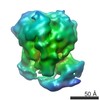
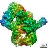
 マップデータ マップデータ | 3D EM map of the specific p53-DNA complex | |||||||||
 試料 試料 |
| |||||||||
 キーワード キーワード | cryo electron microscopy / p53 / single particle analysis / transcription factor | |||||||||
| 生物種 |  | |||||||||
| 手法 | 単粒子再構成法 / クライオ電子顕微鏡法 / 解像度: 21.0 Å | |||||||||
 データ登録者 データ登録者 | Aramayo R / Sherman MB / Brownless K / Lurz R / Okorokov AL / Orlova EV | |||||||||
 引用 引用 | ジャーナル: Nucleic Acids Res / 年: 2011 タイトル: Quaternary structure of the specific p53-DNA complex reveals the mechanism of p53 mutant dominance. 著者: Ricardo Aramayo / Michael B Sherman / Kathryne Brownless / Rudi Lurz / Andrei L Okorokov / Elena V Orlova /  要旨: The p53 tumour suppressor is a transcriptional activator that controls cell fate in response to various stresses. p53 can initiate cell cycle arrest, senescence and/or apoptosis via transactivation ...The p53 tumour suppressor is a transcriptional activator that controls cell fate in response to various stresses. p53 can initiate cell cycle arrest, senescence and/or apoptosis via transactivation of p53 target genes, thus preventing cancer onset. Mutations that impair p53 usually occur in the core domain and negate the p53 sequence-specific DNA binding. Moreover, these mutations exhibit a dominant negative effect on the remaining wild-type p53. Here, we report the cryo electron microscopy structure of the full-length p53 tetramer bound to a DNA-encoding transcription factor response element (RE) at a resolution of 21 A. While two core domains from both dimers of the p53 tetramer interact with DNA within the complex, the other two core domains remain available for binding another DNA site. This finding helps to explain the dominant negative effect of p53 mutants based on the fact that p53 dimers are formed co-translationally before the whole tetramer assembles; therefore, a single mutant dimer would prevent the p53 tetramer from binding DNA. The structure indicates that the Achilles' heel of p53 is in its dimer-of-dimers organization, thus the tetramer activity can be negated by mutation in only one allele followed by tumourigenesis. | |||||||||
| 履歴 |
|
- 構造の表示
構造の表示
| ムービー |
 ムービービューア ムービービューア |
|---|---|
| 構造ビューア | EMマップ: SurfViewMolmilJmol/JSmol |
| 添付画像 |

- ダウンロードとリンク
-EMDBアーカイブ
| マップデータ | emd_1896.map.gz | 395.9 KB | EMDBマップデータ形式 | |
|---|---|---|---|---|
| ヘッダ (付随情報) | emd-1896-v30.xmlemd-1896.xml | 8.6 KB 8.6 KB | 表示 表示 | EMDBヘッダ |
| 画像 | 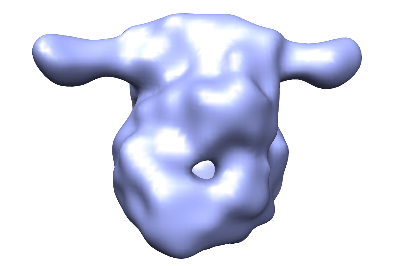 em-1896.png em-1896.png | 248.8 KB | ||
| アーカイブディレクトリ |  http://ftp.pdbj.org/pub/emdb/structures/EMD-1896ftp://ftp.pdbj.org/pub/emdb/structures/EMD-1896 http://ftp.pdbj.org/pub/emdb/structures/EMD-1896ftp://ftp.pdbj.org/pub/emdb/structures/EMD-1896 | HTTPS FTP |
-検証レポート
| 文書・要旨 | emd_1896_validation.pdf.gz | 190.8 KB | 表示 | EMDB検証レポート |
|---|---|---|---|---|
| 文書・詳細版 | emd_1896_full_validation.pdf.gz | 190 KB | 表示 | |
| XML形式データ | emd_1896_validation.xml.gz | 5.3 KB | 表示 | |
| アーカイブディレクトリ | https://ftp.pdbj.org/pub/emdb/validation_reports/EMD-1896ftp://ftp.pdbj.org/pub/emdb/validation_reports/EMD-1896 | HTTPS FTP |
-関連構造データ


-リンク
| EMDBのページ | EMDB (EBI/PDBe) / EMDataResource |
|---|
-マップ
| ファイル | ダウンロード / ファイル: emd_1896.map.gz / 形式: CCP4 / 大きさ: 2.7 MB / タイプ: IMAGE STORED AS FLOATING POINT NUMBER (4 BYTES) | ||||||||||||||||||||||||||||||||||||||||||||||||||||||||||||||||||||
|---|---|---|---|---|---|---|---|---|---|---|---|---|---|---|---|---|---|---|---|---|---|---|---|---|---|---|---|---|---|---|---|---|---|---|---|---|---|---|---|---|---|---|---|---|---|---|---|---|---|---|---|---|---|---|---|---|---|---|---|---|---|---|---|---|---|---|---|---|---|
| 注釈 | 3D EM map of the specific p53-DNA complex | ||||||||||||||||||||||||||||||||||||||||||||||||||||||||||||||||||||
| 投影像・断面図 | 画像のコントロール
画像は Spider により作成 | ||||||||||||||||||||||||||||||||||||||||||||||||||||||||||||||||||||
| ボクセルのサイズ | X=Y=Z: 2.48 Å | ||||||||||||||||||||||||||||||||||||||||||||||||||||||||||||||||||||
| 密度 |
| ||||||||||||||||||||||||||||||||||||||||||||||||||||||||||||||||||||
| 対称性 | 空間群: 1 | ||||||||||||||||||||||||||||||||||||||||||||||||||||||||||||||||||||
| 詳細 | EMDB XML:
CCP4マップ ヘッダ情報:
| ||||||||||||||||||||||||||||||||||||||||||||||||||||||||||||||||||||
 Z (Sec.)
 Y (Row.)
 X (Col.)


-添付データ
- 試料の構成要素
-全体 : Murine p53 tetramer complexed with specific DNA construct
| 全体 | 名称: Murine p53 tetramer complexed with specific DNA construct |
|---|---|
| 要素 |
|
-超分子 #1000: Murine p53 tetramer complexed with specific DNA construct
| 超分子 | 名称: Murine p53 tetramer complexed with specific DNA construct タイプ: sample / ID: 1000 / Number unique components: 2 |
|---|---|
| 分子量 | 実験値: 240 KDa / 理論値: 240 KDa |
-分子 #1: p53
| 分子 | 名称: p53 / タイプ: protein_or_peptide / ID: 1 / Name.synonym: p53 詳細: Murine p53 tetramer bound to a specific DNA construct コピー数: 4 / 集合状態: Tetramer / 組換発現: Yes |
|---|---|
| 由来(天然) | 生物種: |
| 分子量 | 実験値: 240 KDa / 理論値: 240 KDa |
| 組換発現 | 生物種:  unidentified baculovirus (ウイルス) unidentified baculovirus (ウイルス) |
-分子 #2: DNA
| 分子 | 名称: DNA / タイプ: dna / ID: 2 / Name.synonym: DNA / 分類: DNA / Structure: DOUBLE HELIX / Synthetic?: No |
|---|---|
| 由来(天然) | 生物種: synthetic construct (人工物) |
-実験情報
-構造解析
| 手法 | クライオ電子顕微鏡法 |
|---|---|
 解析 解析 | 単粒子再構成法 |
| 試料の集合状態 | particle |
-試料調製
| 緩衝液 | pH: 7 / 詳細: 100mM NaCl, 25mM Tris-HCl, 1mM DTT |
|---|---|
| 凍結 | 凍結剤: ETHANE / チャンバー内温度: 80 K / 装置: OTHER / 手法: Blot few seconds before plunging |
- 電子顕微鏡法
| 顕微鏡 | JEOL 2200FS |
|---|---|
| 温度 | 平均: 90 K |
| 撮影 | カテゴリ: CCD フィルム・検出器のモデル: GATAN ULTRASCAN 4000 (4k x 4k) 平均電子線量: 10 e/Å2 |
| 電子線 | 加速電圧: 200 kV / 電子線源:  FIELD EMISSION GUN FIELD EMISSION GUN |
| 電子光学系 | 照射モード: SPOT SCAN / 撮影モード: BRIGHT FIELD / Cs: 2.0 mm / 最大 デフォーカス(公称値): 5.5 µm / 最小 デフォーカス(公称値): 1.5 µm / 倍率(公称値): 50000 |
| 試料ステージ | 試料ホルダー: Eucentric / 試料ホルダーモデル: GATAN LIQUID NITROGEN |
-画像解析
| CTF補正 | 詳細: Phase flipping |
|---|---|
| 最終 再構成 | 想定した対称性 - 点群: C2 (2回回転対称) / アルゴリズム: OTHER / 解像度のタイプ: BY AUTHOR / 解像度: 21.0 Å / 解像度の算出法: FSC 0.5 CUT-OFF / ソフトウェア - 名称: IMAGIC, EMAN |
