[English] 日本語
 Yorodumi
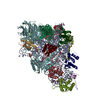
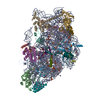
- EMDB-18911: 3DFlex refinement of wheat 40S ribosomal subunit cryo-EM reconstr... -
+ Open data
- Basic information
| Entry |  | |||||||||
|---|---|---|---|---|---|---|---|---|---|---|
| Title | 3DFlex refinement of wheat 40S ribosomal subunit cryo-EM reconstruction | |||||||||
 Map data Map data | ||||||||||
 Sample Sample |
| |||||||||
 Keywords Keywords | small ribosomal subunit / wheat / eukaryotes / 40S / RIBOSOME | |||||||||
| Biological species |  | |||||||||
| Method | single particle reconstruction / cryo EM / Resolution: 2.68 Å | |||||||||
 Authors Authors | Baymukhametov TN / Kravchenko OV / Afonina ZA / Vasilenko KS | |||||||||
| Funding support |  Russian Federation, 1 items Russian Federation, 1 items
| |||||||||
 Citation Citation |  Journal: Int J Mol Sci / Year: 2023 Journal: Int J Mol Sci / Year: 2023Title: High-Resolution Structure and Internal Mobility of a Plant 40S Ribosomal Subunit. Authors: Olesya V Kravchenko / Timur N Baymukhametov / Zhanna A Afonina / Konstantin S Vassilenko /  Abstract: Ribosome is a major part of the protein synthesis machinery, and analysis of its structure is of paramount importance. However, the structure of ribosomes from only a limited number of organisms has ...Ribosome is a major part of the protein synthesis machinery, and analysis of its structure is of paramount importance. However, the structure of ribosomes from only a limited number of organisms has been resolved to date; it especially concerns plant ribosomes and ribosomal subunits. Here, we report a high-resolution cryo-electron microscopy reconstruction of the small subunit of the (common wheat) cytoplasmic ribosome. A detailed atomic model was built that includes the majority of the rRNA and some of the protein modifications. The analysis of the obtained data revealed structural peculiarities of the 40S subunit in the monocot plant ribosome. We applied the 3D Flexible Refinement approach to analyze the internal mobility of the 40S subunit and succeeded in decomposing it into four major motions, describing rotations of the head domain and a shift in the massive rRNA expansion segment. It was shown that these motions are almost uncorrelated and that the 40S subunit is flexible enough to spontaneously adopt any conformation it takes as a part of a translating ribosome or ribosomal complex. Here, we introduce the first high-resolution structure of an isolated plant 40S subunit and the first quantitative analysis of the flexibility of small ribosomal subunits, hoping that it will help in studying various aspects of ribosome functioning. | |||||||||
| History |
|
- Structure visualization
| Supplemental images |
|---|
- Downloads & links
-EMDB archive
| Map data |  emd_18911.map.gz emd_18911.map.gz | 276.6 MB |  EMDB map data format EMDB map data format | |
|---|---|---|---|---|
| Header (meta data) |  emd-18911-v30.xml emd-18911-v30.xml emd-18911.xml emd-18911.xml | 16 KB 16 KB | Display Display |  EMDB header EMDB header |
| FSC (resolution estimation) |  emd_18911_fsc.xml emd_18911_fsc.xml | 17.2 KB | Display |  FSC data file FSC data file |
| Images |  emd_18911.png emd_18911.png | 29.8 KB | ||
| Masks |  emd_18911_msk_1.map emd_18911_msk_1.map | 325 MB |  Mask map Mask map | |
| Filedesc metadata |  emd-18911.cif.gz emd-18911.cif.gz | 4.2 KB | ||
| Others |  emd_18911_half_map_1.map.gz emd_18911_half_map_1.map.gz emd_18911_half_map_2.map.gz emd_18911_half_map_2.map.gz | 16.9 MB 16.9 MB | ||
| Archive directory |  http://ftp.pdbj.org/pub/emdb/structures/EMD-18911 http://ftp.pdbj.org/pub/emdb/structures/EMD-18911 ftp://ftp.pdbj.org/pub/emdb/structures/EMD-18911 ftp://ftp.pdbj.org/pub/emdb/structures/EMD-18911 | HTTPS FTP |
-Related structure data
- Links
Links
| EMDB pages |  EMDB (EBI/PDBe) / EMDB (EBI/PDBe) /  EMDataResource EMDataResource |
|---|
- Map
Map
| File |  Download / File: emd_18911.map.gz / Format: CCP4 / Size: 325 MB / Type: IMAGE STORED AS FLOATING POINT NUMBER (4 BYTES) Download / File: emd_18911.map.gz / Format: CCP4 / Size: 325 MB / Type: IMAGE STORED AS FLOATING POINT NUMBER (4 BYTES) | ||||||||||||||||||||||||||||||||||||
|---|---|---|---|---|---|---|---|---|---|---|---|---|---|---|---|---|---|---|---|---|---|---|---|---|---|---|---|---|---|---|---|---|---|---|---|---|---|
| Projections & slices | Image control
Images are generated by Spider. | ||||||||||||||||||||||||||||||||||||
| Voxel size | X=Y=Z: 0.94 Å | ||||||||||||||||||||||||||||||||||||
| Density |
| ||||||||||||||||||||||||||||||||||||
| Symmetry | Space group: 1 | ||||||||||||||||||||||||||||||||||||
| Details | EMDB XML:
|
-Supplemental data
-Mask #1
| File |  emd_18911_msk_1.map emd_18911_msk_1.map | ||||||||||||
|---|---|---|---|---|---|---|---|---|---|---|---|---|---|
| Projections & Slices |
| ||||||||||||
| Density Histograms |
-Half map: #1
| File | emd_18911_half_map_1.map | ||||||||||||
|---|---|---|---|---|---|---|---|---|---|---|---|---|---|
| Projections & Slices |
| ||||||||||||
| Density Histograms |
-Half map: #2
| File | emd_18911_half_map_2.map | ||||||||||||
|---|---|---|---|---|---|---|---|---|---|---|---|---|---|
| Projections & Slices |
| ||||||||||||
| Density Histograms |
- Sample components
-Entire : Small ribosomal subunit from Triticum aestivum (common wheat)
| Entire | Name: Small ribosomal subunit from Triticum aestivum (common wheat) |
|---|---|
| Components |
|
-Supramolecule #1: Small ribosomal subunit from Triticum aestivum (common wheat)
| Supramolecule | Name: Small ribosomal subunit from Triticum aestivum (common wheat) type: complex / ID: 1 / Parent: 0 / Macromolecule list: #1-#16 |
|---|---|
| Source (natural) | Organism:  |
-Experimental details
-Structure determination
| Method | cryo EM |
|---|---|
 Processing Processing | single particle reconstruction |
| Aggregation state | particle |
- Sample preparation
| Buffer | pH: 7.5 |
|---|---|
| Vitrification | Cryogen name: ETHANE / Chamber humidity: 100 % / Chamber temperature: 283 K / Instrument: FEI VITROBOT MARK IV |
- Electron microscopy
| Microscope | FEI TITAN KRIOS |
|---|---|
| Image recording | Film or detector model: FEI FALCON II (4k x 4k) / Detector mode: INTEGRATING / Digitization - Dimensions - Width: 4096 pixel / Digitization - Dimensions - Height: 4096 pixel / Digitization - Frames/image: 0-32 / Number grids imaged: 1 / Number real images: 6201 / Average exposure time: 1.6 sec. / Average electron dose: 84.0 e/Å2 |
| Electron beam | Acceleration voltage: 300 kV / Electron source:  FIELD EMISSION GUN FIELD EMISSION GUN |
| Electron optics | C2 aperture diameter: 100.0 µm / Illumination mode: FLOOD BEAM / Imaging mode: BRIGHT FIELD / Cs: 0.1 mm / Nominal defocus max: 1.8 µm / Nominal defocus min: 0.6 µm / Nominal magnification: 75000 |
| Sample stage | Specimen holder model: FEI TITAN KRIOS AUTOGRID HOLDER / Cooling holder cryogen: NITROGEN |
| Experimental equipment |  Model: Titan Krios / Image courtesy: FEI Company |
 Movie
Movie Controller


 Z (Sec.)
 Y (Row.)
 X (Col.)


