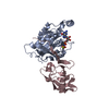
ジャーナル: Nat Struct Mol Biol / 年: 2025 タイトル: Capture, mutual inhibition and release mechanism for aPKC-Par6 and its multisite polarity substrate Lgl. 著者: Christopher P Earl / Mathias Cobbaut / André Barros-Carvalho / Marina E Ivanova / David C Briggs / Eurico Morais-de-Sá / Peter J Parker / Neil Q McDonald / 要旨: The mutually antagonistic relationship of atypical protein kinase C (aPKC) and partitioning-defective protein 6 (Par6) with the substrate lethal (2) giant larvae (Lgl) is essential for regulating ...The mutually antagonistic relationship of atypical protein kinase C (aPKC) and partitioning-defective protein 6 (Par6) with the substrate lethal (2) giant larvae (Lgl) is essential for regulating polarity across many cell types. Although aPKC-Par6 phosphorylates Lgl at three serine sites to exclude it from the apical domain, aPKC-Par6 and Lgl paradoxically form a stable kinase-substrate complex, with conflicting roles proposed for Par6. We report the structure of human aPKCι-Par6α bound to full-length Llgl1, captured through an aPKCι docking site and a Par6 contact. This complex traps a phospho-S663 Llgl1 intermediate bridging between aPKC and Par6, impeding phosphorylation progression. Thus, aPKCι is effectively inhibited by Llgl1 while Llgl1 is captured by aPKCι-Par6. Mutational disruption of the Lgl-aPKC interaction impedes complex assembly and Lgl phosphorylation, whereas disrupting the Lgl-Par6 contact promotes complex dissociation and Lgl phosphorylation. We demonstrate a Par6-regulated substrate capture-and-release model requiring binding by active Cdc42 and the apical partner Crumbs to drive complex disassembly. Our results suggest a mechanism for mutual regulation and spatial control of aPKC-Par6 and Lgl activities.
凍結剤: ETHANE / チャンバー内湿度: 100 % / チャンバー内温度: 298 K / 装置: FEI VITROBOT MARK IV 詳細: 4 ul of aPKCiota-Par6-Llgl1 complex at a concentration of 0.4 mg/ml was applied to R1.2/1.3 Quantifoil 300 mesh copper grids which had been glow-discharged for 45 s at 45 mA . Grids were ...詳細: 4 ul of aPKCiota-Par6-Llgl1 complex at a concentration of 0.4 mg/ml was applied to R1.2/1.3 Quantifoil 300 mesh copper grids which had been glow-discharged for 45 s at 45 mA . Grids were blotted for 2.5 s at 100% humidity using an FEI Vitrobot MK IV..
詳細
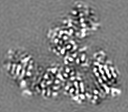
Sample was double affinity purified and gel filtered. Sample was monodisperse.
-
電子顕微鏡法
顕微鏡
FEI TITAN KRIOS
特殊光学系
エネルギーフィルター - 名称: GIF Quantum ER
撮影
フィルム・検出器のモデル: GATAN K2 SUMMIT (4k x 4k) 検出モード: COUNTING / 実像数: 4002 / 平均露光時間: 8.0 sec. / 平均電子線量: 48.1 e/Å2
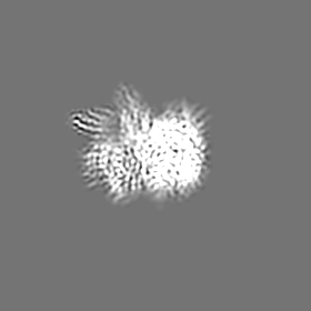
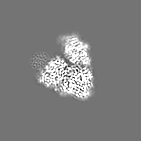
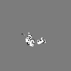
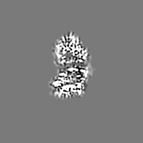
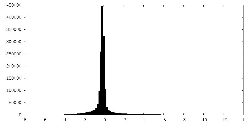
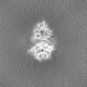
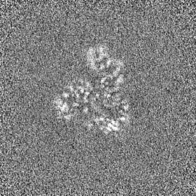
選択した数: 1069057 詳細: Semi-automated picking with Xmipp3 and particle extraction in Relion-3 yielded 47,516 particles from 1000 micrographs 38. After reference-free 2D classification in Relion-3, eight 2D classes ...詳細: Semi-automated picking with Xmipp3 and particle extraction in Relion-3 yielded 47,516 particles from 1000 micrographs 38. After reference-free 2D classification in Relion-3, eight 2D classes were selected and used as templates for reference-based particle picking in Gautomatch. A total of particles were extracted with 2-fold binning and submitted to 8 rounds of 2D classification in Relion-3.
 ムービー
ムービー コントローラー


 データを開く
 基本情報
基本情報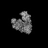
 マップデータ
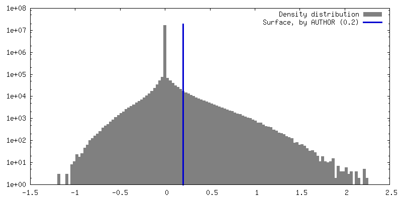
 試料
試料 キーワード
キーワード 機能・相同性情報
 Homo sapiens (ヒト)
 データ登録者
 英国, 3件
  引用
引用
 構造の表示
構造の表示
 ダウンロードとリンク
 emd_18877.png
 http://ftp.pdbj.org/pub/emdb/structures/EMD-18877


 Z (Sec.)
 X (Row.)
 Y (Col.)


 試料の構成要素
 解析
解析 電子顕微鏡法
 FIELD EMISSION GUN