 Movie
Movie Controller


+ Open data

- Basic information
| Entry |  | ||||||||||||||||||
|---|---|---|---|---|---|---|---|---|---|---|---|---|---|---|---|---|---|---|---|
| Title | wild type neuronal synapse | ||||||||||||||||||
 Map data Map data | wild type neuronal synapse. non filtered map. | ||||||||||||||||||
 Sample Sample |
| ||||||||||||||||||
 Keywords Keywords | synapse / neurotransmission / EXOCYTOSIS | ||||||||||||||||||
| Biological species |  | ||||||||||||||||||
| Method | electron tomography / cryo EM | ||||||||||||||||||
 Authors Authors | Radecke J / Seeger R / Zuber B / Sorensen JB | ||||||||||||||||||
| Funding support |  Switzerland, Switzerland,  Denmark, 5 items Denmark, 5 items
| ||||||||||||||||||
 Citation Citation | Journal: EMBO Rep / Year: 2023 Title: Morphofunctional changes at the active zone during synaptic vesicle exocytosis. Authors: Julika Radecke / Raphaela Seeger / Anna Kádková / Ulrike Laugks / Amin Khosrozadeh / Kenneth N Goldie / Vladan Lučić / Jakob B Sørensen / Benoît Zuber /   Abstract: Synaptic vesicle (SV) fusion with the plasma membrane (PM) proceeds through intermediate steps that remain poorly resolved. The effect of persistent high or low exocytosis activity on intermediate ...Synaptic vesicle (SV) fusion with the plasma membrane (PM) proceeds through intermediate steps that remain poorly resolved. The effect of persistent high or low exocytosis activity on intermediate steps remains unknown. Using spray-mixing plunge-freezing cryo-electron tomography we observe events following synaptic stimulation at nanometer resolution in near-native samples. Our data suggest that during the stage that immediately follows stimulation, termed early fusion, PM and SV membrane curvature changes to establish a point contact. The next stage-late fusion-shows fusion pore opening and SV collapse. During early fusion, proximal tethered SVs form additional tethers with the PM and increase the inter-SV connector number. In the late-fusion stage, PM-proximal SVs lose their interconnections, allowing them to move toward the PM. Two SNAP-25 mutations, one arresting and one disinhibiting spontaneous release, cause connector loss. The disinhibiting mutation causes loss of membrane-proximal multiple-tethered SVs. Overall, tether formation and connector dissolution are triggered by stimulation and respond to spontaneous fusion rate manipulation. These morphological observations likely correspond to SV transition from one functional pool to another. | ||||||||||||||||||
| History |
|
- Structure visualization
| Supplemental images |
|---|

- Downloads & links
-EMDB archive
| Map data | emd_18746.map.gz | 310.4 MB |  EMDB map data format EMDB map data format | |
|---|---|---|---|---|
| Header (meta data) | emd-18746-v30.xmlemd-18746.xml | 12.6 KB 12.6 KB | Display Display | EMDB header |
| Images | 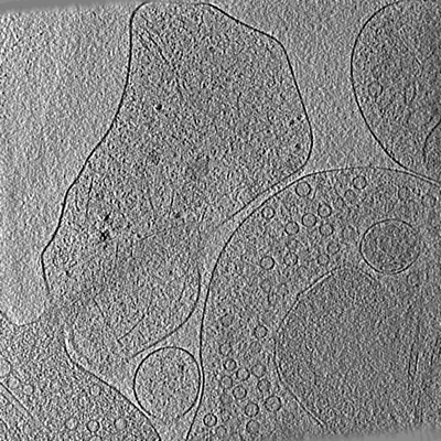 emd_18746.png emd_18746.png | 287 KB | ||
| Filedesc metadata | emd-18746.cif.gz | 4.3 KB | ||
| Others | emd_18746_additional_1.map.gz | 253.4 MB | ||
| Archive directory |  http://ftp.pdbj.org/pub/emdb/structures/EMD-18746ftp://ftp.pdbj.org/pub/emdb/structures/EMD-18746 http://ftp.pdbj.org/pub/emdb/structures/EMD-18746ftp://ftp.pdbj.org/pub/emdb/structures/EMD-18746 | HTTPS FTP |
-Related structure data


-Links
| EMDB pages | EMDB (EBI/PDBe) / EMDataResource |
|---|
-Map
| File | Download / File: emd_18746.map.gz / Format: CCP4 / Size: 608 MB / Type: IMAGE STORED AS SIGNED INTEGER (2 BYTES) | ||||||||||||||||||||||||||||||||
|---|---|---|---|---|---|---|---|---|---|---|---|---|---|---|---|---|---|---|---|---|---|---|---|---|---|---|---|---|---|---|---|---|---|
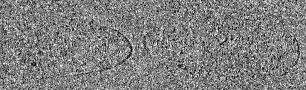
| Annotation | wild type neuronal synapse. non filtered map. | ||||||||||||||||||||||||||||||||
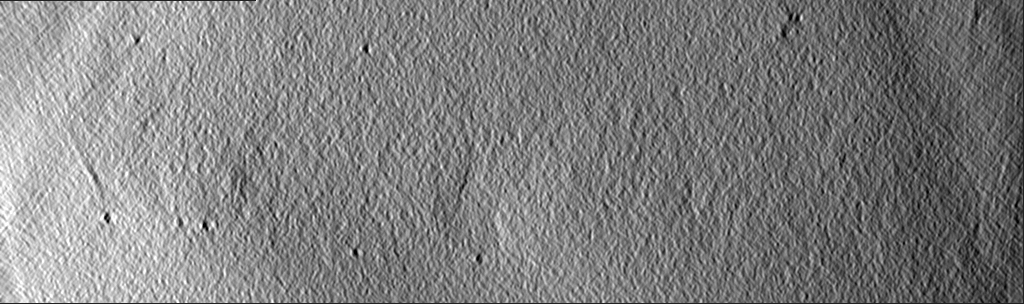
| Projections & slices | Image control
Images are generated by Spider. generated in cubic-lattice coordinate | ||||||||||||||||||||||||||||||||
| Voxel size | X=Y=Z: 14.69 Å | ||||||||||||||||||||||||||||||||
| Density |
| ||||||||||||||||||||||||||||||||
| Symmetry | Space group: 1 | ||||||||||||||||||||||||||||||||
| Details | EMDB XML:
|
 Z (Sec.)
 Y (Row.)
 X (Col.)


-Supplemental data
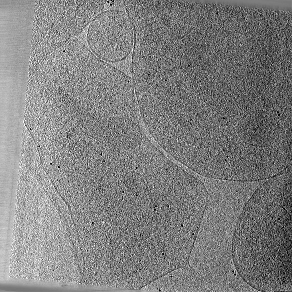
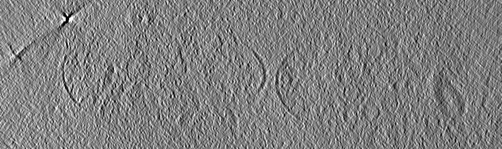
-Additional map: wild type neuronal synapse. Non-linear anistropic diffusion filtered...
| File | emd_18746_additional_1.map | ||||||||||||
|---|---|---|---|---|---|---|---|---|---|---|---|---|---|
| Annotation | wild type neuronal synapse. Non-linear anistropic diffusion filtered map | ||||||||||||
| Projections & Slices |
| ||||||||||||
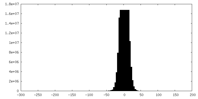
| Density Histograms |


- Sample components
-Entire : Synapse between hippocampal neurons isolated from SNAP-25 KO and ...
| Entire | Name: Synapse between hippocampal neurons isolated from SNAP-25 KO and transduced with SNAP-25 wild type. |
|---|---|
| Components |
|
-Supramolecule #1: Synapse between hippocampal neurons isolated from SNAP-25 KO and ...
| Supramolecule | Name: Synapse between hippocampal neurons isolated from SNAP-25 KO and transduced with SNAP-25 wild type. type: cell / ID: 1 / Parent: 0 |
|---|---|
| Source (natural) | Organism: |
-Experimental details
-Structure determination
| Method | cryo EM |
|---|---|
 Processing Processing | electron tomography |
| Aggregation state | cell |
-Sample preparation
| Buffer | pH: 7.4 Details: NB medium (Neurobasal with 2% B-27, 1 M HEPES, 0.26% lutamax, 14.3 mM beta-mercaptoethanol, 10000 IU penicillin, 10 mg streptomycin) |
|---|---|
| Grid | Model: Quantifoil R2/2 / Material: GOLD / Support film - Material: CARBON / Support film - topology: HOLEY ARRAY |
| Vitrification | Cryogen name: ETHANE / Chamber humidity: 100 % / Chamber temperature: 277 K / Instrument: FEI VITROBOT MARK IV Details: After 12 to 14 days of incubation grids with mouse neurons were plunge frozen with a Vitrobot (Thermofisher Scientific, Mark IV) with a blot time of 3 s and a blot force of -10. Wait time ...Details: After 12 to 14 days of incubation grids with mouse neurons were plunge frozen with a Vitrobot (Thermofisher Scientific, Mark IV) with a blot time of 3 s and a blot force of -10. Wait time and drain time were not used. Humidity was set to 100% at 4 degrees C. 4 ul undiluted 10 nm BSA gold tracer (Aurion) was added directly onto the grid prior to plunge freezing.. |
| Sectioning | Other: NO SECTIONING |
| Fiducial marker | Manufacturer: AURION Immuno Gold Reagents & Accessories / Diameter: 10 nm |
- Electron microscopy
| Microscope | FEI TITAN KRIOS |
|---|---|
| Image recording | Film or detector model: FEI FALCON III (4k x 4k) / Detector mode: INTEGRATING / Average electron dose: 0.76 e/Å2 |
| Electron beam | Acceleration voltage: 300 kV / Electron source:  FIELD EMISSION GUN FIELD EMISSION GUN |
| Electron optics | Illumination mode: FLOOD BEAM / Imaging mode: BRIGHT FIELD / Nominal defocus max: 10.0 µm / Nominal defocus min: 6.0 µm |
| Sample stage | Specimen holder model: FEI TITAN KRIOS AUTOGRID HOLDER / Cooling holder cryogen: NITROGEN |
| Experimental equipment |  Model: Titan Krios / Image courtesy: FEI Company |
-Image processing
| Final reconstruction | Algorithm: SIMULTANEOUS ITERATIVE (SIRT) / Software - Name: IMOD / Number images used: 131 |
|---|
