Journal: EMBO Rep / Year: 2023 Title: Morphofunctional changes at the active zone during synaptic vesicle exocytosis. Authors: Julika Radecke / Raphaela Seeger / Anna Kádková / Ulrike Laugks / Amin Khosrozadeh / Kenneth N Goldie / Vladan Lučić / Jakob B Sørensen / Benoît Zuber / Abstract: Synaptic vesicle (SV) fusion with the plasma membrane (PM) proceeds through intermediate steps that remain poorly resolved. The effect of persistent high or low exocytosis activity on intermediate ...Synaptic vesicle (SV) fusion with the plasma membrane (PM) proceeds through intermediate steps that remain poorly resolved. The effect of persistent high or low exocytosis activity on intermediate steps remains unknown. Using spray-mixing plunge-freezing cryo-electron tomography we observe events following synaptic stimulation at nanometer resolution in near-native samples. Our data suggest that during the stage that immediately follows stimulation, termed early fusion, PM and SV membrane curvature changes to establish a point contact. The next stage-late fusion-shows fusion pore opening and SV collapse. During early fusion, proximal tethered SVs form additional tethers with the PM and increase the inter-SV connector number. In the late-fusion stage, PM-proximal SVs lose their interconnections, allowing them to move toward the PM. Two SNAP-25 mutations, one arresting and one disinhibiting spontaneous release, cause connector loss. The disinhibiting mutation causes loss of membrane-proximal multiple-tethered SVs. Overall, tether formation and connector dissolution are triggered by stimulation and respond to spontaneous fusion rate manipulation. These morphological observations likely correspond to SV transition from one functional pool to another.
A: 12902.399 Å / B: 14694.399 Å / C: 3942.4 Å α=β=γ: 90.0 °
-
Supplemental data
-
Sample components
-
Entire : Stimulated rat synaptosome
Entire
Name: Stimulated rat synaptosome
Components
Cell: Stimulated rat synaptosome
-
Supramolecule #1: Stimulated rat synaptosome
Supramolecule
Name: Stimulated rat synaptosome / type: cell / ID: 1 / Parent: 0 Details: sprayed with KCl milliseconds before freezing in order to stimulate exocytosis.
In the structure databanks used in Yorodumi, some data are registered as the other names, "COVID-19 virus" and "2019-nCoV". Here are the details of the virus and the list of structure data.
Jan 31, 2019. EMDB accession codes are about to change! (news from PDBe EMDB page)
EMDB accession codes are about to change! (news from PDBe EMDB page)
The allocation of 4 digits for EMDB accession codes will soon come to an end. Whilst these codes will remain in use, new EMDB accession codes will include an additional digit and will expand incrementally as the available range of codes is exhausted. The current 4-digit format prefixed with “EMD-” (i.e. EMD-XXXX) will advance to a 5-digit format (i.e. EMD-XXXXX), and so on. It is currently estimated that the 4-digit codes will be depleted around Spring 2019, at which point the 5-digit format will come into force.
The EM Navigator/Yorodumi systems omit the EMD- prefix.
Related info.:Q: What is EMD? / ID/Accession-code notation in Yorodumi/EM Navigator
Yorodumi is a browser for structure data from EMDB, PDB, SASBDB, etc.
This page is also the successor to EM Navigator detail page, and also detail information page/front-end page for Omokage search.
The word "yorodu" (or yorozu) is an old Japanese word meaning "ten thousand". "mi" (miru) is to see.
Related info.:EMDB / PDB / SASBDB / Comparison of 3 databanks / Yorodumi Search / Aug 31, 2016. New EM Navigator & Yorodumi / Yorodumi Papers / Jmol/JSmol / Function and homology information / Changes in new EM Navigator and Yorodumi
 Movie
Movie Controller


 Open data
 Basic information
 Map data
 Sample
 Keywords
 Authors
 Switzerland, 1 items
  Citation


 Structure visualization
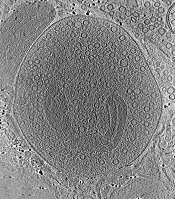
 Downloads & links
 EMDB map data format
 emd_18744.png
 http://ftp.pdbj.org/pub/emdb/structures/EMD-18744


 Z (Sec.)
 Y (Row.)
 X (Col.)


 Sample components
 Processing
 Electron microscopy
 FIELD EMISSION GUN