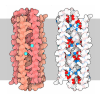
+ データを開く
- 基本情報
基本情報
| 登録情報 |  | |||||||||
|---|---|---|---|---|---|---|---|---|---|---|
| タイトル | Cryo-EM structure of the heat-irreversible amyloid fibrils of human lysozyme | |||||||||
 マップデータ マップデータ | ||||||||||
 試料 試料 |
| |||||||||
 キーワード キーワード | amyloid / irreversible / PROTEIN FIBRIL | |||||||||
| 機能・相同性 |  機能・相同性情報 機能・相同性情報antimicrobial humoral response / Antimicrobial peptides / specific granule lumen / azurophil granule lumen / lysozyme / lysozyme activity / tertiary granule lumen / defense response to Gram-negative bacterium / killing of cells of another organism / defense response to Gram-positive bacterium ...antimicrobial humoral response / Antimicrobial peptides / specific granule lumen / azurophil granule lumen / lysozyme / lysozyme activity / tertiary granule lumen / defense response to Gram-negative bacterium / killing of cells of another organism / defense response to Gram-positive bacterium / defense response to bacterium / inflammatory response / Amyloid fiber formation / Neutrophil degranulation / extracellular space / extracellular exosome / extracellular region / identical protein binding 類似検索 - 分子機能 | |||||||||
| 生物種 |  Homo sapiens (ヒト) Homo sapiens (ヒト) | |||||||||
| 手法 | らせん対称体再構成法 / クライオ電子顕微鏡法 / 解像度: 2.8 Å | |||||||||
 データ登録者 データ登録者 | Frey L / Greenwald J / Riek R | |||||||||
| 資金援助 |  スイス, 1件 スイス, 1件
| |||||||||
 引用 引用 |  ジャーナル: Nat Commun / 年: 2024 ジャーナル: Nat Commun / 年: 2024タイトル: A structural rationale for reversible vs irreversible amyloid fibril formation from a single protein. 著者: Lukas Frey / Jiangtao Zhou / Gea Cereghetti / Marco E Weber / David Rhyner / Aditya Pokharna / Luca Wenchel / Harindranath Kadavath / Yiping Cao / Beat H Meier / Matthias Peter / Jason ...著者: Lukas Frey / Jiangtao Zhou / Gea Cereghetti / Marco E Weber / David Rhyner / Aditya Pokharna / Luca Wenchel / Harindranath Kadavath / Yiping Cao / Beat H Meier / Matthias Peter / Jason Greenwald / Roland Riek / Raffaele Mezzenga /    要旨: Reversible and irreversible amyloids are two diverging cases of protein (mis)folding associated with the cross-β motif in the protein folding and aggregation energy landscape. Yet, the molecular ...Reversible and irreversible amyloids are two diverging cases of protein (mis)folding associated with the cross-β motif in the protein folding and aggregation energy landscape. Yet, the molecular origins responsible for the formation of reversible vs irreversible amyloids have remained unknown. Here we provide evidence at the atomic level of distinct folding motifs for irreversible and reversible amyloids derived from a single protein sequence: human lysozyme. We compare the 2.8 Å structure of irreversible amyloid fibrils determined by cryo-electron microscopy helical reconstructions with molecular insights gained by solid-state NMR spectroscopy on reversible amyloids. We observe a canonical cross-β-sheet structure in irreversible amyloids, whereas in reversible amyloids, there is a less-ordered coexistence of β-sheet and helical secondary structures that originate from a partially unfolded lysozyme, thus carrying a "memory" of the original folded protein precursor. We also report the structure of hen egg-white lysozyme irreversible amyloids at 3.2 Å resolution, revealing another canonical amyloid fold, and reaffirming that irreversible amyloids undergo a complete conversion of the native protein into the cross-β structure. By combining atomic force microscopy, cryo-electron microscopy and solid-state NMR, we show that a full unfolding of the native protein precursor is a requirement for establishing irreversible amyloid fibrils. | |||||||||
| 履歴 |
|
- 構造の表示
構造の表示
- ダウンロードとリンク
-EMDBアーカイブ
| マップデータ |  emd_18663.map.gz emd_18663.map.gz | 4.8 MB |  EMDBマップデータ形式 EMDBマップデータ形式 | |
|---|---|---|---|---|
| ヘッダ (付随情報) |  emd-18663-v30.xml emd-18663-v30.xml emd-18663.xml emd-18663.xml | 15.9 KB 15.9 KB | 表示 表示 |  EMDBヘッダ EMDBヘッダ |
| FSC (解像度算出) |  emd_18663_fsc.xml emd_18663_fsc.xml | 9.1 KB | 表示 |  FSCデータファイル FSCデータファイル |
| 画像 |  emd_18663.png emd_18663.png | 68.1 KB | ||
| Filedesc metadata |  emd-18663.cif.gz emd-18663.cif.gz | 5.6 KB | ||
| その他 |  emd_18663_half_map_1.map.gz emd_18663_half_map_1.map.gz emd_18663_half_map_2.map.gz emd_18663_half_map_2.map.gz | 49.6 MB 49.6 MB | ||
| アーカイブディレクトリ |  http://ftp.pdbj.org/pub/emdb/structures/EMD-18663 http://ftp.pdbj.org/pub/emdb/structures/EMD-18663 ftp://ftp.pdbj.org/pub/emdb/structures/EMD-18663 ftp://ftp.pdbj.org/pub/emdb/structures/EMD-18663 | HTTPS FTP |
-検証レポート
| 文書・要旨 |  emd_18663_validation.pdf.gz emd_18663_validation.pdf.gz | 665.2 KB | 表示 |  EMDB検証レポート EMDB検証レポート |
|---|---|---|---|---|
| 文書・詳細版 |  emd_18663_full_validation.pdf.gz emd_18663_full_validation.pdf.gz | 664.8 KB | 表示 | |
| XML形式データ |  emd_18663_validation.xml.gz emd_18663_validation.xml.gz | 15.5 KB | 表示 | |
| CIF形式データ |  emd_18663_validation.cif.gz emd_18663_validation.cif.gz | 20.2 KB | 表示 | |
| アーカイブディレクトリ |  https://ftp.pdbj.org/pub/emdb/validation_reports/EMD-18663 https://ftp.pdbj.org/pub/emdb/validation_reports/EMD-18663 ftp://ftp.pdbj.org/pub/emdb/validation_reports/EMD-18663 ftp://ftp.pdbj.org/pub/emdb/validation_reports/EMD-18663 | HTTPS FTP |
-関連構造データ
| 関連構造データ |  8qutMC  8qv8C M: このマップから作成された原子モデル C: 同じ文献を引用 ( |
|---|---|
| 類似構造データ | 類似検索 - 機能・相同性  F&H 検索 F&H 検索 |
- リンク
リンク
| EMDBのページ |  EMDB (EBI/PDBe) / EMDB (EBI/PDBe) /  EMDataResource EMDataResource |
|---|---|
| 「今月の分子」の関連する項目 |
- マップ
マップ
| ファイル |  ダウンロード / ファイル: emd_18663.map.gz / 形式: CCP4 / 大きさ: 64 MB / タイプ: IMAGE STORED AS FLOATING POINT NUMBER (4 BYTES) ダウンロード / ファイル: emd_18663.map.gz / 形式: CCP4 / 大きさ: 64 MB / タイプ: IMAGE STORED AS FLOATING POINT NUMBER (4 BYTES) | ||||||||||||||||||||||||||||||||||||
|---|---|---|---|---|---|---|---|---|---|---|---|---|---|---|---|---|---|---|---|---|---|---|---|---|---|---|---|---|---|---|---|---|---|---|---|---|---|
| 投影像・断面図 | 画像のコントロール
画像は Spider により作成 | ||||||||||||||||||||||||||||||||||||
| ボクセルのサイズ | X=Y=Z: 1.3 Å | ||||||||||||||||||||||||||||||||||||
| 密度 |
| ||||||||||||||||||||||||||||||||||||
| 対称性 | 空間群: 1 | ||||||||||||||||||||||||||||||||||||
| 詳細 | EMDB XML:
|
-添付データ
- 試料の構成要素
-全体 : lysozyme amyloid fibril
| 全体 | 名称: lysozyme amyloid fibril |
|---|---|
| 要素 |
|
-超分子 #1: lysozyme amyloid fibril
| 超分子 | 名称: lysozyme amyloid fibril / タイプ: complex / ID: 1 / 親要素: 0 / 含まれる分子: all |
|---|---|
| 由来(天然) | 生物種:  Homo sapiens (ヒト) Homo sapiens (ヒト) |
-分子 #1: Lysozyme C
| 分子 | 名称: Lysozyme C / タイプ: protein_or_peptide / ID: 1 / コピー数: 5 / 光学異性体: LEVO |
|---|---|
| 由来(天然) | 生物種:  Homo sapiens (ヒト) Homo sapiens (ヒト) |
| 分子量 | 理論値: 14.720693 KDa |
| 組換発現 | 生物種:  |
| 配列 | 文字列: KVFERCELAR TLKRLGMDGY RGISLANWMC LAKWESGYNT RATNYNAGDR STDYGIFQIN SRYWCNDGKT PGAVNACHLS CSALLQDNI ADAVACAKRV VRDPQGIRAW VAWRNRCQNR DVRQYVQGCG V UniProtKB: Lysozyme C |
-分子 #2: Unidentified peptide
| 分子 | 名称: Unidentified peptide / タイプ: protein_or_peptide / ID: 2 / コピー数: 5 / 光学異性体: LEVO |
|---|---|
| 由来(天然) | 生物種:  Homo sapiens (ヒト) Homo sapiens (ヒト) |
| 分子量 | 理論値: 869.063 Da |
| 組換発現 | 生物種:  |
| 配列 | 文字列: (UNK)(UNK)(UNK)(UNK)(UNK)(UNK)(UNK)(UNK)(UNK)(UNK) |
-実験情報
-構造解析
| 手法 | クライオ電子顕微鏡法 |
|---|---|
 解析 解析 | らせん対称体再構成法 |
| 試料の集合状態 | filament |
- 試料調製
試料調製
| 緩衝液 | pH: 7 / 詳細: 20mM DTT |
|---|---|
| 凍結 | 凍結剤: ETHANE-PROPANE |
- 電子顕微鏡法
| 顕微鏡 | FEI TITAN KRIOS |
|---|---|
| 特殊光学系 | エネルギーフィルター - 名称: GIF Bioquantum / エネルギーフィルター - スリット幅: 20 eV |
| 撮影 | フィルム・検出器のモデル: GATAN K3 BIOQUANTUM (6k x 4k) 実像数: 926 / 平均露光時間: 1.0 sec. / 平均電子線量: 62.79 e/Å2 |
| 電子線 | 加速電圧: 300 kV / 電子線源:  FIELD EMISSION GUN FIELD EMISSION GUN |
| 電子光学系 | C2レンズ絞り径: 70.0 µm / 照射モード: FLOOD BEAM / 撮影モード: BRIGHT FIELD / Cs: 2.7 mm / 最大 デフォーカス(公称値): 3.0 µm / 最小 デフォーカス(公称値): 0.8 µm / 倍率(公称値): 130000 |
| 試料ステージ | 試料ホルダーモデル: FEI TITAN KRIOS AUTOGRID HOLDER ホルダー冷却材: NITROGEN |
| 実験機器 |  モデル: Titan Krios / 画像提供: FEI Company |
- 画像解析
画像解析
-原子モデル構築 1
| 精密化 | 空間: REAL / プロトコル: AB INITIO MODEL |
|---|---|
| 得られたモデル |  PDB-8qut: |
 ムービー
ムービー コントローラー


 Z (Sec.)
 Y (Row.)
 X (Col.)


